Copyright
©The Author(s) 2020.
World J Clin Cases. Dec 26, 2020; 8(24): 6296-6305
Published online Dec 26, 2020. doi: 10.12998/wjcc.v8.i24.6296
Published online Dec 26, 2020. doi: 10.12998/wjcc.v8.i24.6296
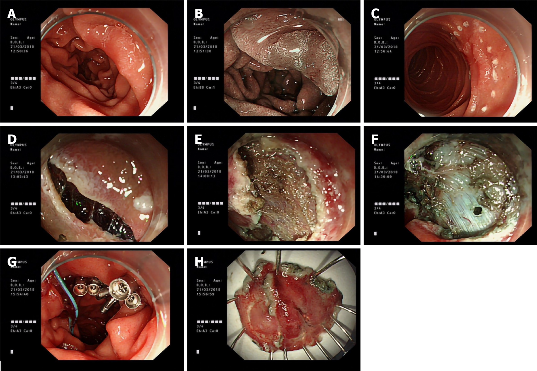
Figure 2 Endoscopic submucosal dissection was performed for a depressed lesion in the descending duodenum.
A flat polypoid elevation approximately 15 mm in diameter with central depression was observed in the duodenal bulb, hybrid endoscopic submucosal dissection was performed, and pathology suggested a tubular adenoma. A: A depressed lesion in the descending duodenum; B: NBI showed that the lesion boundary was clear; C: Mark the lesion boundary; D: The mucosa layer was incised circumferentiality; E: Dissect the lesions along the submucosa; F: Muscular perforation; G: Cover the perforation with nylon rope and metal clips; H: Resected specimen for pathological examination.
- Citation: Li XY, Ji KY, Qu YH, Zheng JJ, Guo YJ, Zhang CP, Zhang KP. Application of endoscopic submucosal dissection in duodenal space-occupying lesions. World J Clin Cases 2020; 8(24): 6296-6305
- URL: https://www.wjgnet.com/2307-8960/full/v8/i24/6296.htm
- DOI: https://dx.doi.org/10.12998/wjcc.v8.i24.6296


