Copyright
©The Author(s) 2020.
World J Clin Cases. Dec 6, 2020; 8(23): 6190-6196
Published online Dec 6, 2020. doi: 10.12998/wjcc.v8.i23.6190
Published online Dec 6, 2020. doi: 10.12998/wjcc.v8.i23.6190
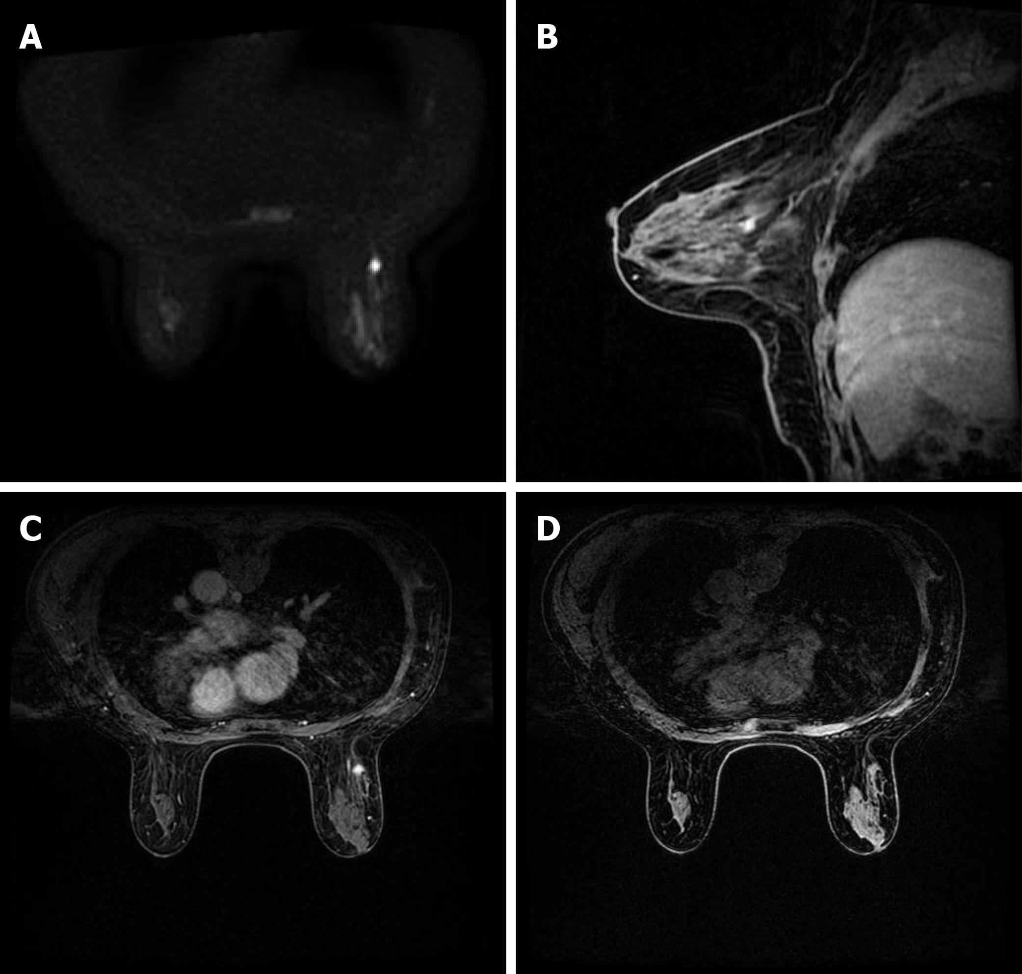
Figure 2 Brest imaging.
A: Axial diffusion weighted imaging (b = 800) showed the mass with high signal intensity; B and C: Sagittal and axial post-contrast fat-saturated T1-weighted magnetic resonance imaging identified a 0.8 × 0.9 cm enhancing irregular mass at the 9-10 o'clock position, approximately 6 cm from the nipple; D: Transverse non-enhanced T1 weighted imaging revealed an ill-defined mass with uniform equal signal intensity.
- Citation: Wang XM, Cong YZ, Qiao GD, Zhang S, Wang LJ. Primary breast cancer patient with poliomyelitis: A case report. World J Clin Cases 2020; 8(23): 6190-6196
- URL: https://www.wjgnet.com/2307-8960/full/v8/i23/6190.htm
- DOI: https://dx.doi.org/10.12998/wjcc.v8.i23.6190


