Copyright
©The Author(s) 2020.
World J Clin Cases. Dec 6, 2020; 8(23): 6190-6196
Published online Dec 6, 2020. doi: 10.12998/wjcc.v8.i23.6190
Published online Dec 6, 2020. doi: 10.12998/wjcc.v8.i23.6190
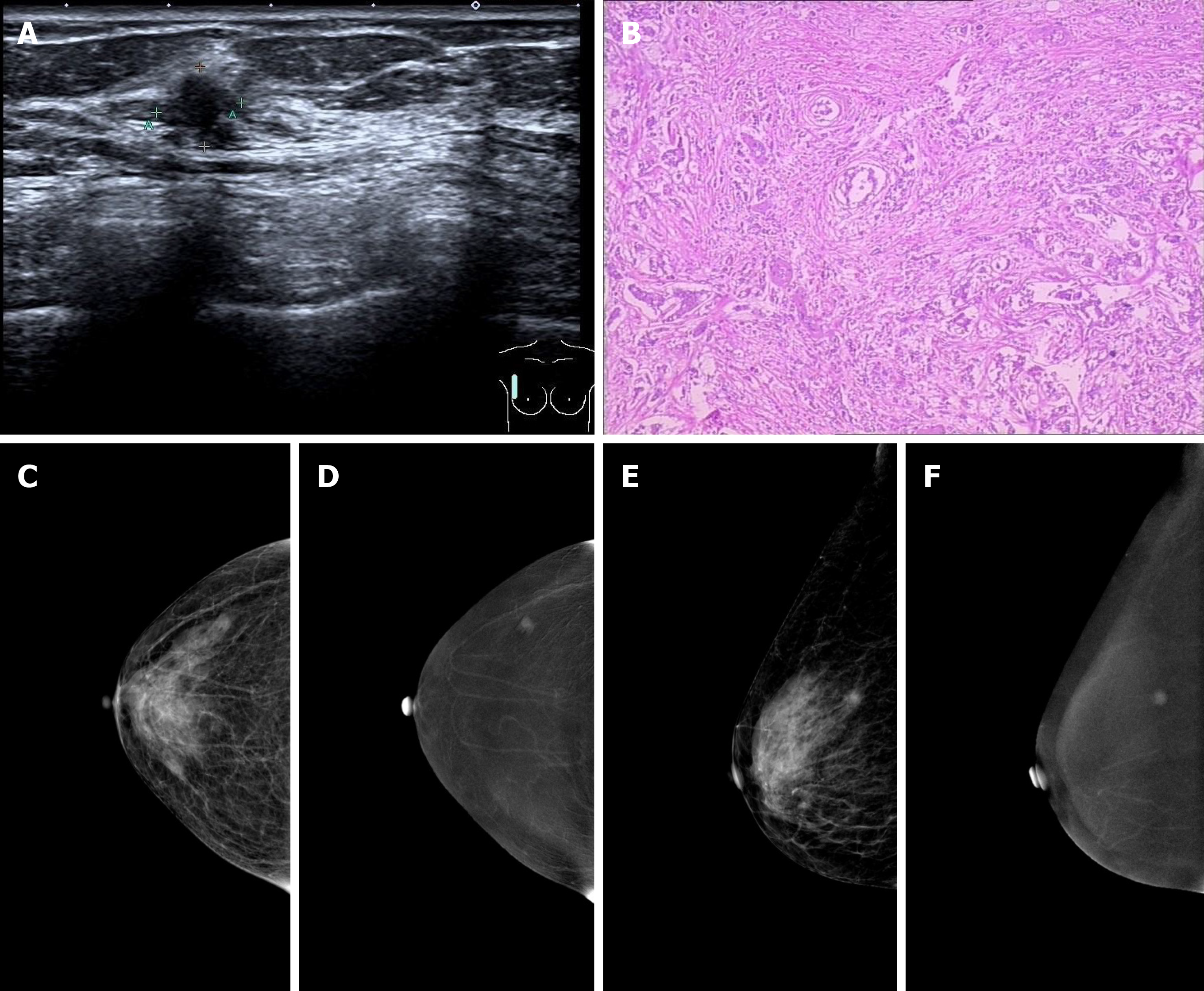
Figure 1 Breast imaging.
A: Ultrasound examination revealed an ill-defined hypoechoic mass in the right breast with an irregular shape; B: Microscopic examination revealed invasive ductal carcinoma cells invading the parenchyma in the right breast; C and E: On contrast enhanced spectral mammography (CESM) imaging, the low energy images showed the mass on the upper outer quadrant of the right breast with small spicules at the periphery; D and F: On CESM imaging, the subtraction images showed the significant enhancement of the lesions.
- Citation: Wang XM, Cong YZ, Qiao GD, Zhang S, Wang LJ. Primary breast cancer patient with poliomyelitis: A case report. World J Clin Cases 2020; 8(23): 6190-6196
- URL: https://www.wjgnet.com/2307-8960/full/v8/i23/6190.htm
- DOI: https://dx.doi.org/10.12998/wjcc.v8.i23.6190


