Copyright
©The Author(s) 2020.
World J Clin Cases. Dec 6, 2020; 8(23): 6172-6180
Published online Dec 6, 2020. doi: 10.12998/wjcc.v8.i23.6172
Published online Dec 6, 2020. doi: 10.12998/wjcc.v8.i23.6172
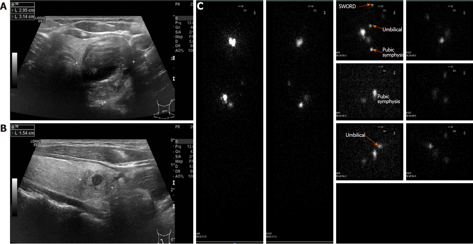
Figure 3 Ultrasound and 99mTc sodium pertechnetate scintigraphy of the thyroid.
A-B: Ultrasound of the thyroid showed nodular goiters in both normal lateral lobes; C: 99mTc sodium pertechnetate scintigraphy showed three abnormal foci in the left renal and lower abdomen.
- Citation: Qin LH, He FY, Liao JY. Multiple ectopic goiter in the retroperitoneum, abdominal wall, liver, and diaphragm: A case report and review of literature. World J Clin Cases 2020; 8(23): 6172-6180
- URL: https://www.wjgnet.com/2307-8960/full/v8/i23/6172.htm
- DOI: https://dx.doi.org/10.12998/wjcc.v8.i23.6172


