Copyright
©The Author(s) 2020.
World J Clin Cases. Dec 6, 2020; 8(23): 6144-6149
Published online Dec 6, 2020. doi: 10.12998/wjcc.v8.i23.6144
Published online Dec 6, 2020. doi: 10.12998/wjcc.v8.i23.6144
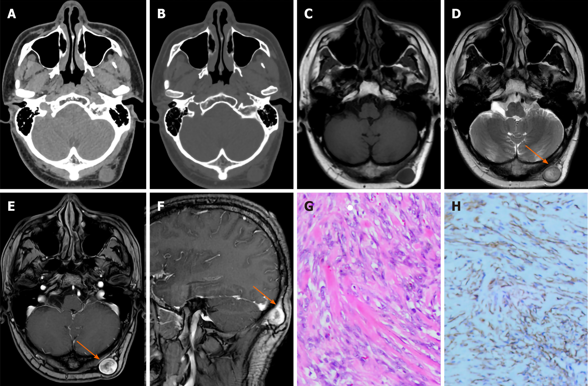
Figure 1 Imaging examination and immunohistochemistry images.
A: Computed tomography showed a subcutaneous nodule with a diameter of approximately 2 cm in the left occipital area, with uniform density, clear boundaries, and smooth edges; B: No destruction of the bone adjacent to the skull; C: Cross-sectional T1-weighted imaging (T1WI) showed that the superficial fascia close to the left occipital showed a round iso-signal nodule spreading to the subcutaneous fat layer, and the edge was clear and smooth; D: Cross-sectional T2-weighted imaging (T2WI) showed that the lesion had inhomogeneous high signal intensity, and the central signal was higher than that of the periphery (arrow); E: Cross-sectional contrast-enhanced T1WI showed that the lesion had obvious inhomogeneous enhancement, with a higher degree of peripheral enhancement than that of the center, showing an "inverted target sign" (arrow); F: Sagittal contrast-enhanced T1WI showed obvious fascia thickening and enhancement adjacent to the focus, showing a "fascia tail sign" (arrow); G: A large number of proliferated spindle cells were arranged erratically and mucoid matrix, local angiogenesis, and a small amount of erythrocyte extravasation could be seen (hematoxylin-eosin staining, × 200); H: Positive expression of SMA in spindle cells in the lesion (immunohistochemical staining, × 200).
- Citation: Wang T, Tang GC, Yang H, Fan JK. Occipital nodular fasciitis easily misdiagnosed as neoplastic lesions: A rare case report. World J Clin Cases 2020; 8(23): 6144-6149
- URL: https://www.wjgnet.com/2307-8960/full/v8/i23/6144.htm
- DOI: https://dx.doi.org/10.12998/wjcc.v8.i23.6144


