Copyright
©The Author(s) 2020.
World J Clin Cases. Dec 6, 2020; 8(23): 6136-6143
Published online Dec 6, 2020. doi: 10.12998/wjcc.v8.i23.6136
Published online Dec 6, 2020. doi: 10.12998/wjcc.v8.i23.6136
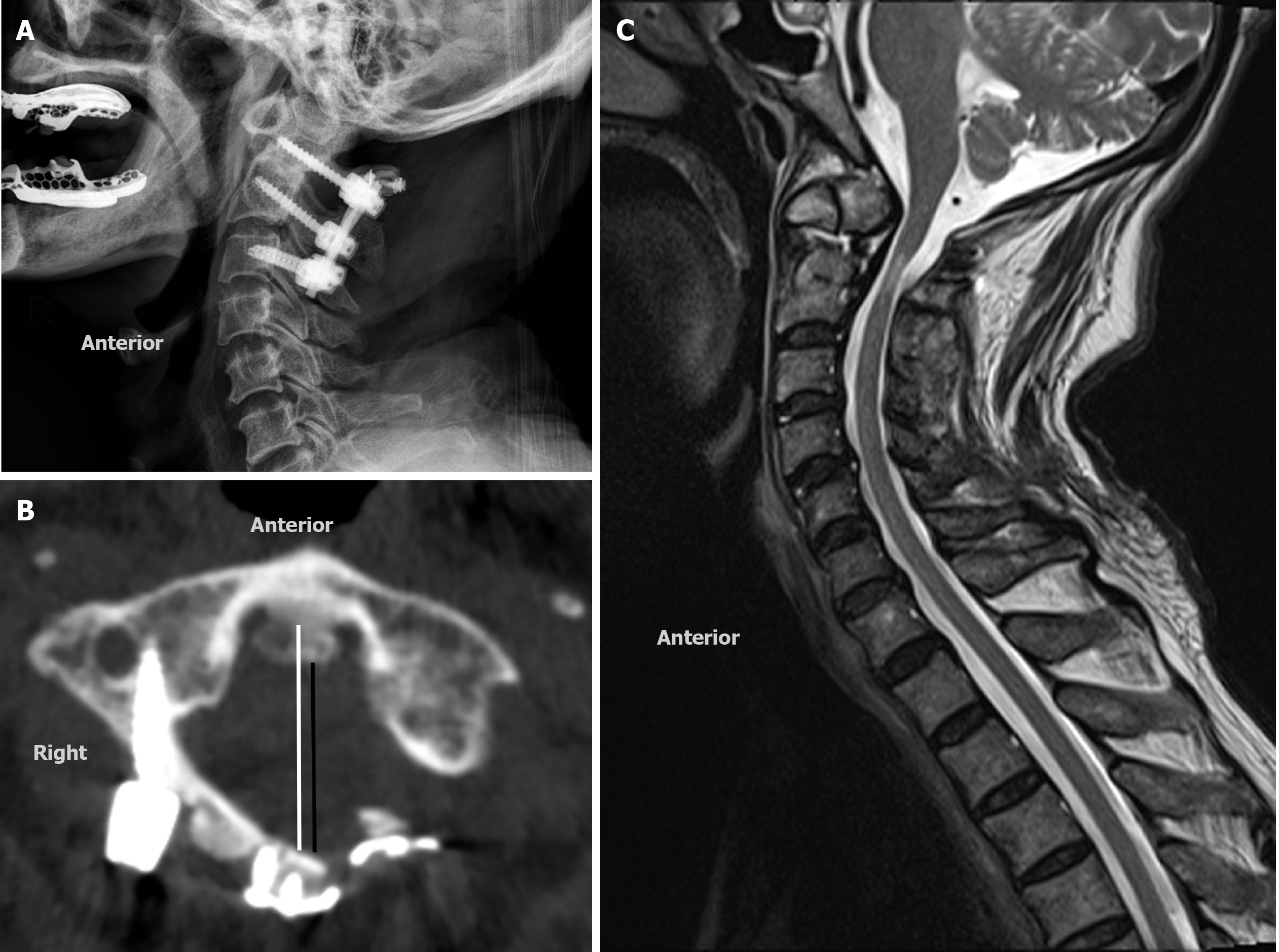
Figure 3 Postoperative images during the 3-yr follow up.
A: Lateral radiograph indicating stable fixation and fusion; B: Axial computed tomography scan, demonstrating the C1 inner sagittal diameter (white line) = 32.75 mm and the canal sagittal diameter (black line) = 22.24 mm; C: Parasagittal magnetic resonance imaging, demonstrating satisfactory release of compression caused by the odontoid process and C1 posterior arch.
- Citation: Zhu Y, Wu XX, Jiang AQ, Li XF, Yang HL, Jiang WM. Single door laminoplasty plus posterior fusion for posterior atlantoaxial dislocation with congenital malformation: A case report and review of literature. World J Clin Cases 2020; 8(23): 6136-6143
- URL: https://www.wjgnet.com/2307-8960/full/v8/i23/6136.htm
- DOI: https://dx.doi.org/10.12998/wjcc.v8.i23.6136


