Copyright
©The Author(s) 2020.
World J Clin Cases. Dec 6, 2020; 8(23): 6136-6143
Published online Dec 6, 2020. doi: 10.12998/wjcc.v8.i23.6136
Published online Dec 6, 2020. doi: 10.12998/wjcc.v8.i23.6136
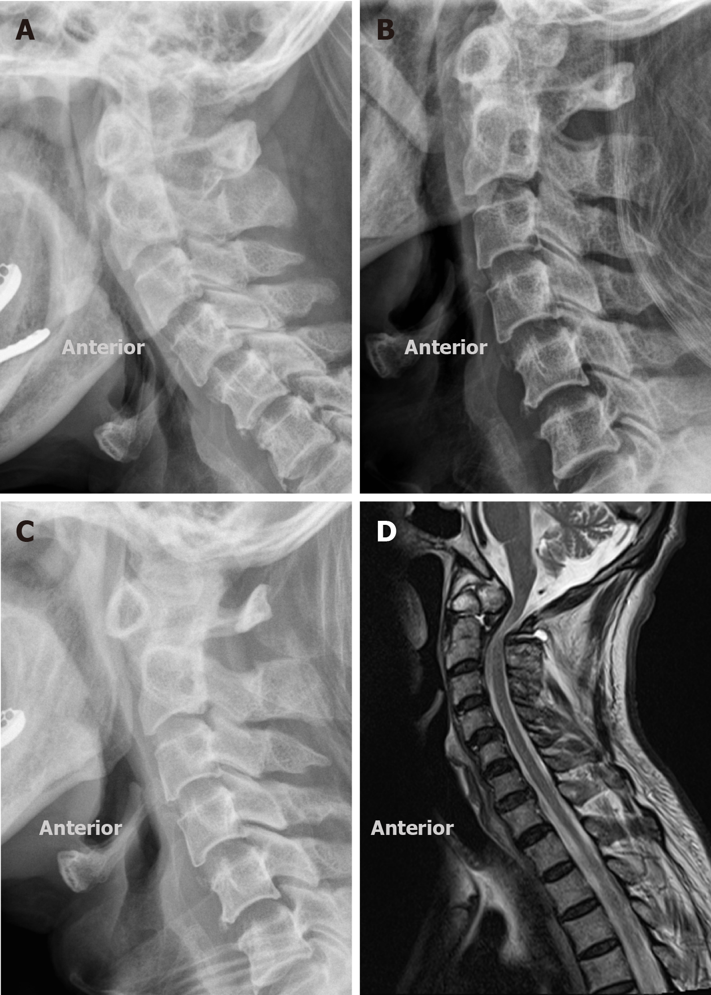
Figure 2 Images of reduction.
A: Lateral radiographs 4 d after traction; B: Lateral radiographs 8 d after traction; C: Lateral radiographs 12 d after traction, showing satisfactory closed reduction; D: Post-reductional parasagittal magnetic resonance imaging, demonstrating that the spinal cord was still compressed by C1 posterior arch.
- Citation: Zhu Y, Wu XX, Jiang AQ, Li XF, Yang HL, Jiang WM. Single door laminoplasty plus posterior fusion for posterior atlantoaxial dislocation with congenital malformation: A case report and review of literature. World J Clin Cases 2020; 8(23): 6136-6143
- URL: https://www.wjgnet.com/2307-8960/full/v8/i23/6136.htm
- DOI: https://dx.doi.org/10.12998/wjcc.v8.i23.6136


