Copyright
©The Author(s) 2020.
World J Clin Cases. Dec 6, 2020; 8(23): 6110-6121
Published online Dec 6, 2020. doi: 10.12998/wjcc.v8.i23.6110
Published online Dec 6, 2020. doi: 10.12998/wjcc.v8.i23.6110
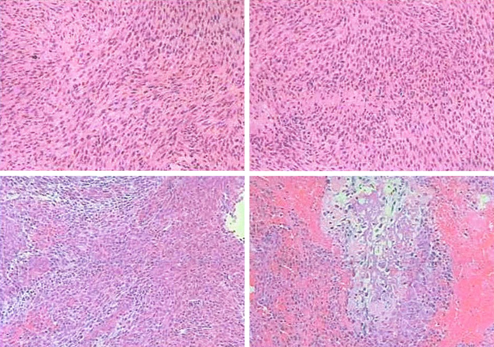
Figure 4 Microscopic pathology.
A spindle cell malignant tumor with local osteogenesis; and vimentin (+ ), CK (-), EMA (+), CK5 / 6 (-), P40 (-), CD34 (-), S-100 (-), SMA (+), TTF-1 (-), calponin [small focus] (+), desmin (-), PR (-), D2-40 (-), E-CAD (-), and Ki-67 [+ (50%)]. In situ hybridization showed EBERs (-). The following diagnoses were considered: (1) Osteosarcoma; (2) Malignant meningiomas with heterogeneous differentiation; (3) Tumor tissue noted in the common carotid artery; (4) Tumor tissue noted in the brachiocephalic trunk; and (5) One tracheoesophageal lymph node without metastasis.
- Citation: Chen HY, Zhao F, Qin JY, Lin HM, Su JP. Malignant meningioma with jugular vein invasion and carotid artery extension: A case report and review of the literature. World J Clin Cases 2020; 8(23): 6110-6121
- URL: https://www.wjgnet.com/2307-8960/full/v8/i23/6110.htm
- DOI: https://dx.doi.org/10.12998/wjcc.v8.i23.6110


