Copyright
©The Author(s) 2020.
World J Clin Cases. Dec 6, 2020; 8(23): 6110-6121
Published online Dec 6, 2020. doi: 10.12998/wjcc.v8.i23.6110
Published online Dec 6, 2020. doi: 10.12998/wjcc.v8.i23.6110
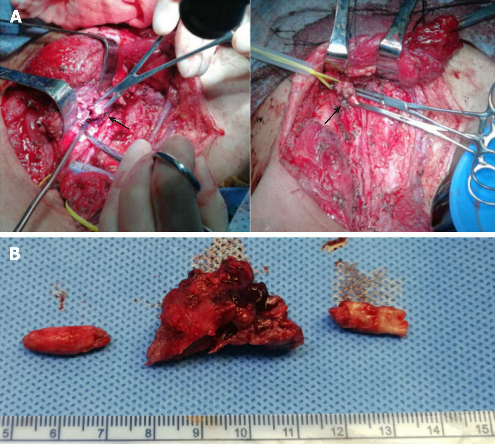
Figure 3 Surgical findings.
A: Shows a tumor thrombus in the right common carotid artery (left black arrow). Tumor invasion at the junction of the common carotid artery and the subclavian artery was also observed (right black arrow); B: Shows the removed tumor thrombus. The tumor thrombus in the artery is columnar in shape, the width of which is consistent with the inner diameter of the blood vessel, and there is a large amount of calcified tissue in the tumor.
- Citation: Chen HY, Zhao F, Qin JY, Lin HM, Su JP. Malignant meningioma with jugular vein invasion and carotid artery extension: A case report and review of the literature. World J Clin Cases 2020; 8(23): 6110-6121
- URL: https://www.wjgnet.com/2307-8960/full/v8/i23/6110.htm
- DOI: https://dx.doi.org/10.12998/wjcc.v8.i23.6110


