Copyright
©The Author(s) 2020.
World J Clin Cases. Dec 6, 2020; 8(23): 6110-6121
Published online Dec 6, 2020. doi: 10.12998/wjcc.v8.i23.6110
Published online Dec 6, 2020. doi: 10.12998/wjcc.v8.i23.6110
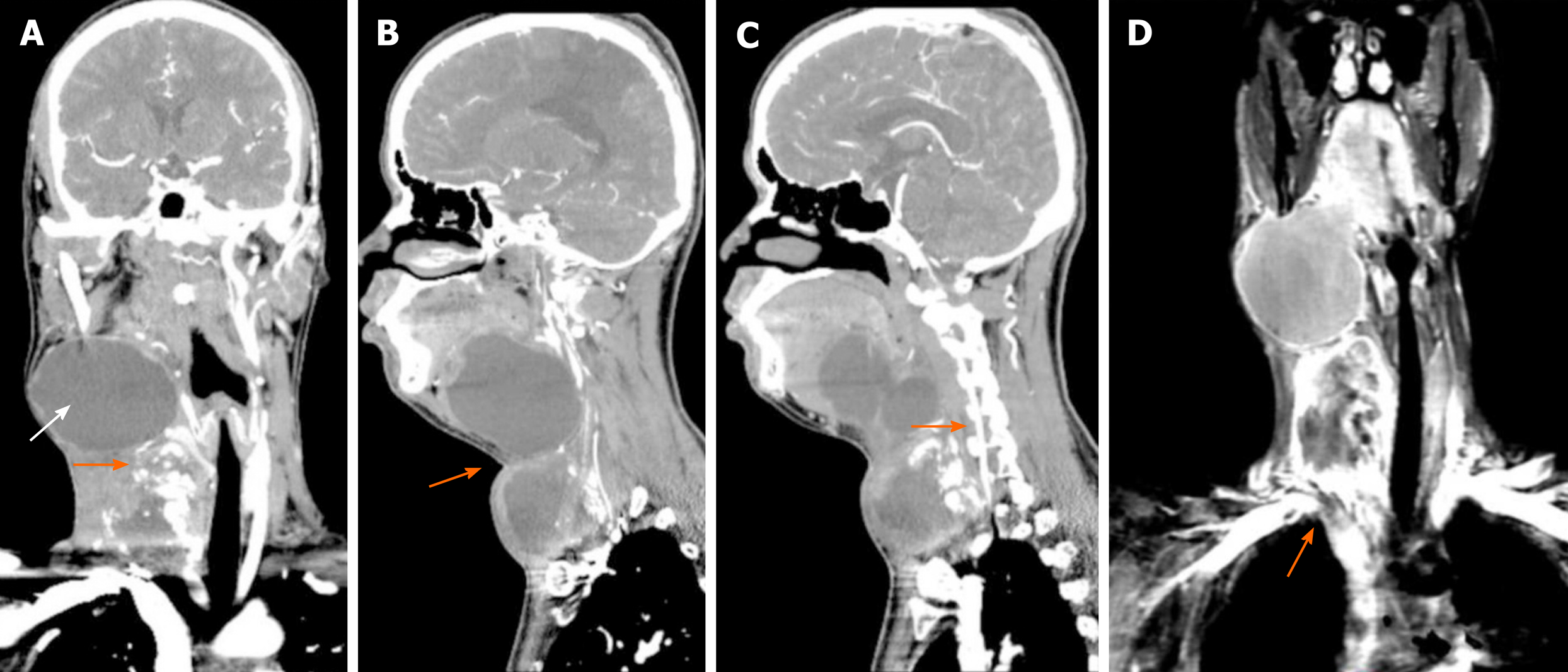
Figure 2 Computed tomography and magnetic resonance imaging.
A: Computed tomography imaging revealed a right side calcified mass and an unclear boundary at the root of the neck measuring 7.1 cm × 5.8 cm × 6.0 cm (orange arrow). Another large cystic mass was present in the right parapharyngeal space and measured 5.9 cm × 9.2 cm × 8.1 cm (white arrow). The two masses reached the right sublingual space and extended downward to the thorax. The adjacent blood vessels were displaced and the trachea, oropharynx, and hypopharynx were deviated to the left; B: The boundary between the upper and lower cervical masses is blurred (orange arrow); C: The vertebral artery is very close to the tumor (orange arrow). The vertebral artery runs inside the cervical spine. If the vertebral artery is injured during surgical resection of the tumor, it is likely to cause death due to the difficulty of hemostasis; D: Magnetic resonance imaging revealed a discontinuity of the angiography at the junction of the right subclavian artery (orange arrow) and the common carotid artery, indicating that the tumor has invaded the right subclavian artery.
- Citation: Chen HY, Zhao F, Qin JY, Lin HM, Su JP. Malignant meningioma with jugular vein invasion and carotid artery extension: A case report and review of the literature. World J Clin Cases 2020; 8(23): 6110-6121
- URL: https://www.wjgnet.com/2307-8960/full/v8/i23/6110.htm
- DOI: https://dx.doi.org/10.12998/wjcc.v8.i23.6110


