Copyright
©The Author(s) 2020.
World J Clin Cases. Dec 6, 2020; 8(23): 6080-6085
Published online Dec 6, 2020. doi: 10.12998/wjcc.v8.i23.6080
Published online Dec 6, 2020. doi: 10.12998/wjcc.v8.i23.6080
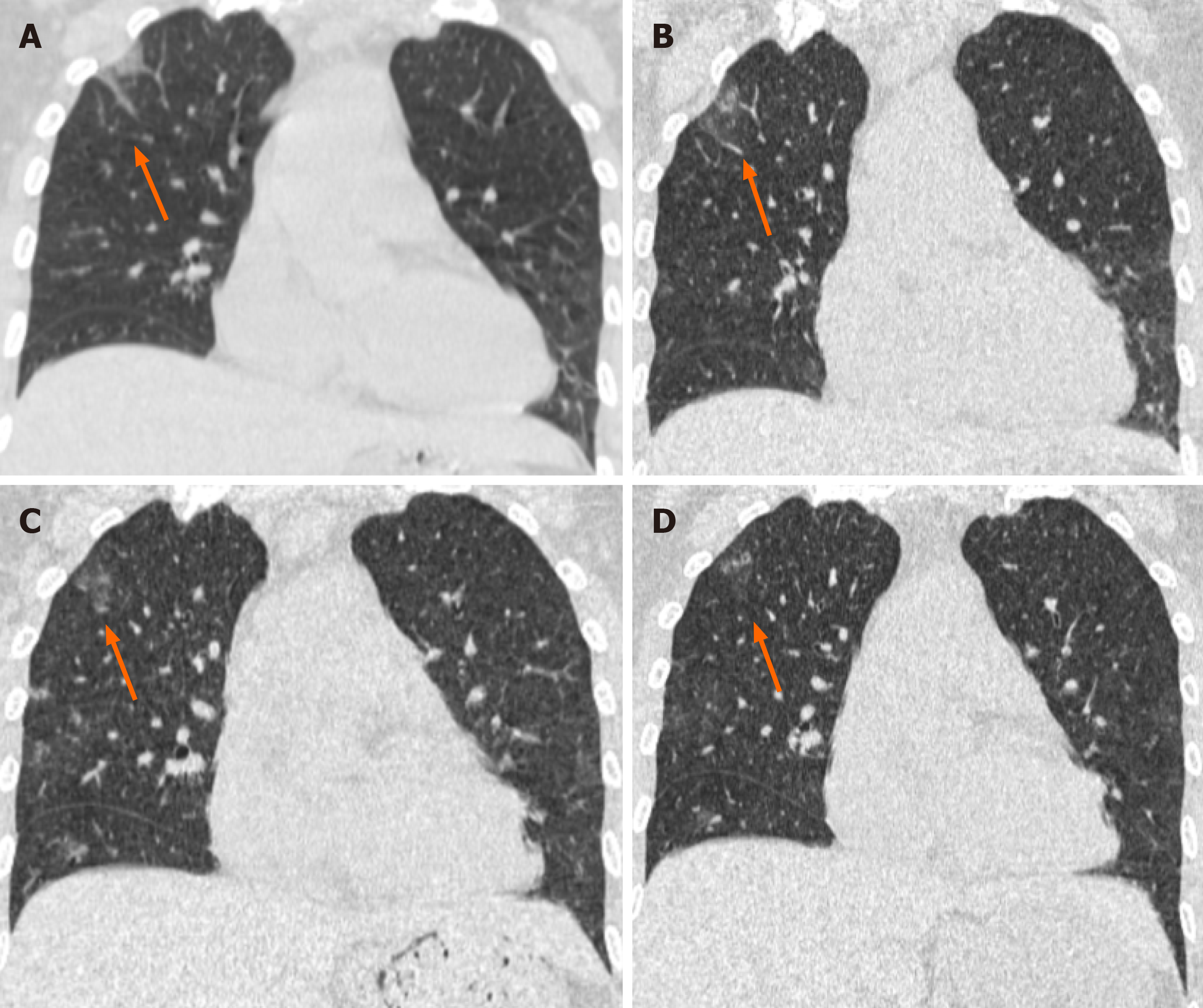
Figure 2 Unenhanced computed tomography images in a 49-year-old woman (coronal imaging).
Computed tomography radiographs show a patchy and striate shadow of slightly high density in the upper right lung (arrows), as the treatment progressed, this asymmetrical lesion was gradually absorbed. A: Day 7 (2020.1.26) after the onset of symptoms; B: Day 12 (2020.1.31) after the onset of symptoms; C: Day 16 (2020.2.4) after the onset of symptoms; D: Day 21 (2020.2.9) after the onset of symptoms.
- Citation: Gao ZA, Gao LB, Chen XJ, Xu Y. Fourty-nine years old woman co-infected with SARS-CoV-2 and Mycoplasma: A case report. World J Clin Cases 2020; 8(23): 6080-6085
- URL: https://www.wjgnet.com/2307-8960/full/v8/i23/6080.htm
- DOI: https://dx.doi.org/10.12998/wjcc.v8.i23.6080


