Copyright
©The Author(s) 2020.
World J Clin Cases. Dec 6, 2020; 8(23): 6036-6042
Published online Dec 6, 2020. doi: 10.12998/wjcc.v8.i23.6036
Published online Dec 6, 2020. doi: 10.12998/wjcc.v8.i23.6036
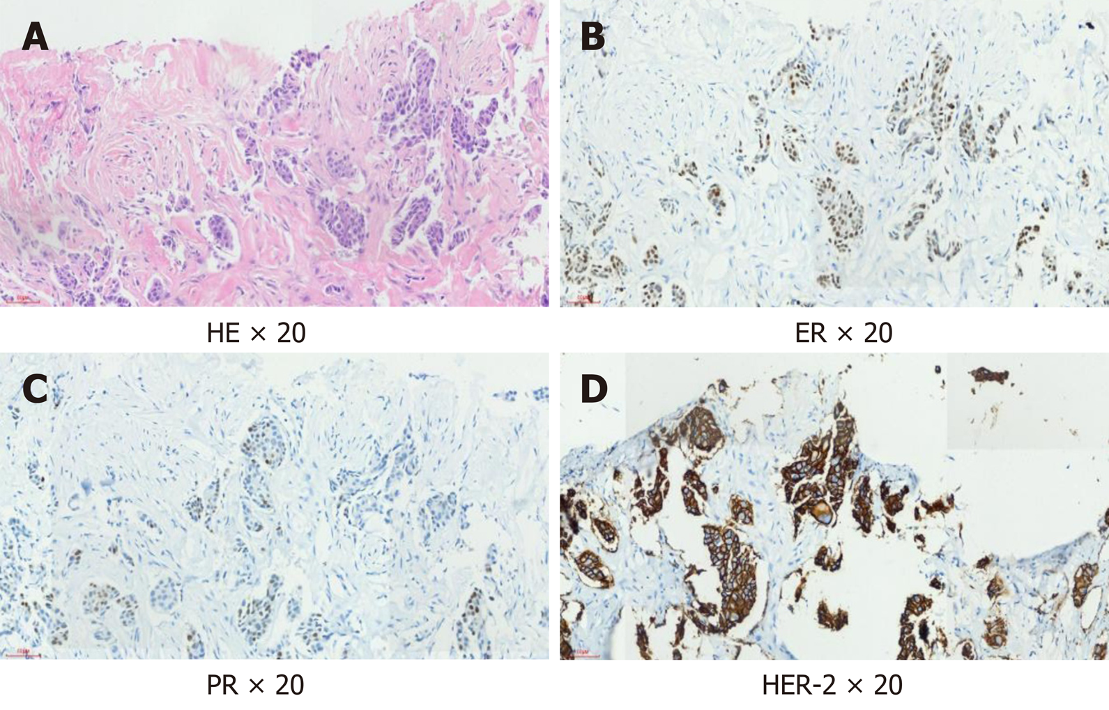
Figure 1 Immunohistochemical examination of the left breast mass.
A: Hematoxylin-eosin staining, 20 ×; B: Estrogen receptor, 20 ×; C: Progesterone receptor, 20 ×; D: Human epidermal factor receptor 2, 20 ×. HE: Hematoxylin-eosin; ER: Estrogen receptor; PR: Progesterone receptor; HER2: Human epidermal factor receptor 2.
- Citation: Yang P, Peng SJ, Dong YM, Yang L, Yang ZY, Hu XE, Bao GQ. Neoadjuvant targeted therapy for apocrine carcinoma of the breast: A case report. World J Clin Cases 2020; 8(23): 6036-6042
- URL: https://www.wjgnet.com/2307-8960/full/v8/i23/6036.htm
- DOI: https://dx.doi.org/10.12998/wjcc.v8.i23.6036


