Copyright
©The Author(s) 2020.
World J Clin Cases. Dec 6, 2020; 8(23): 6009-6015
Published online Dec 6, 2020. doi: 10.12998/wjcc.v8.i23.6009
Published online Dec 6, 2020. doi: 10.12998/wjcc.v8.i23.6009
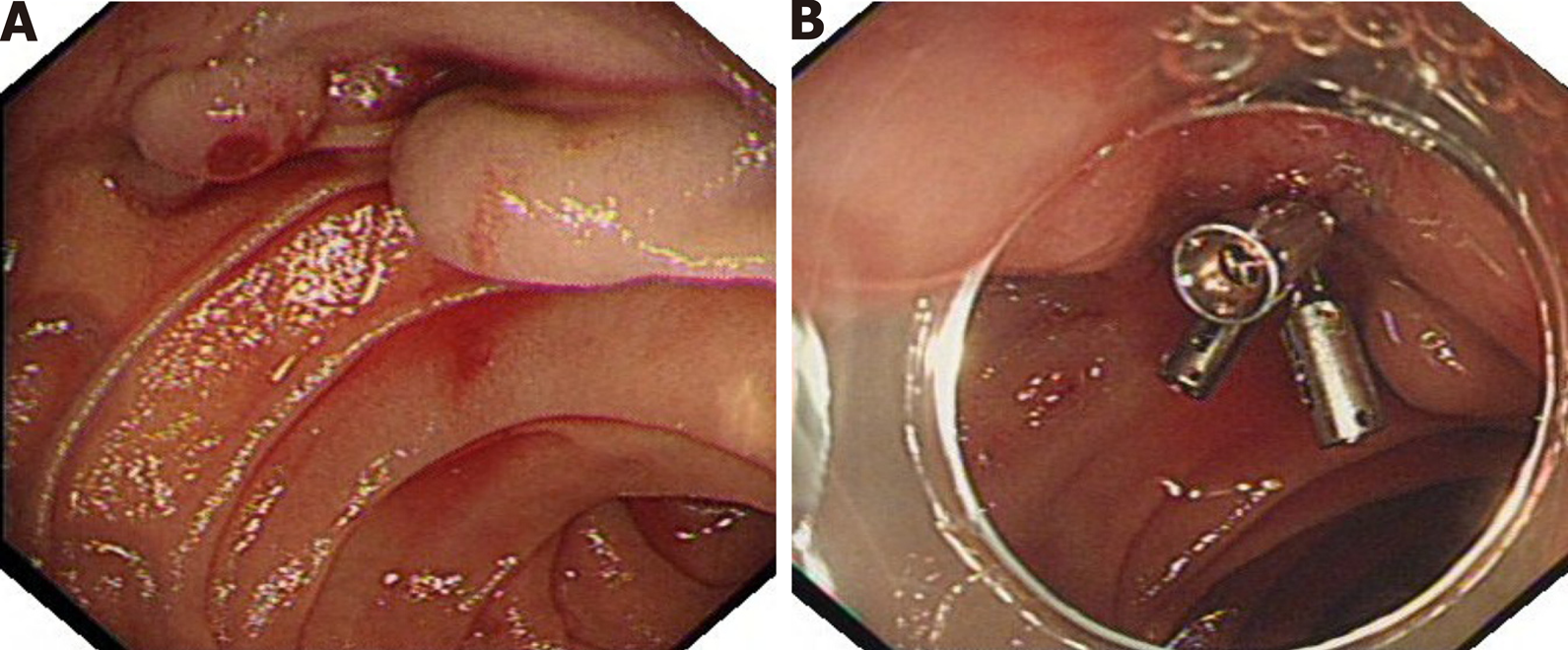
Figure 1 The first endoscopic examination found that the patient had duodenal variceal bleeding.
A: A plurality of tortuous gray-blue varicose veins are seen in the horizontal part of duodenum, with the thickest diameter of about 0.7 cm, active bleeding can be seen on the surface of varicose veins; B: The surface of varicose veins is clamped by metal clips, and bleeding stops.
- Citation: Li DH, Liu XY, Xu LB. Duodenal giant stromal tumor combined with ectopic varicose hemorrhage: A case report. World J Clin Cases 2020; 8(23): 6009-6015
- URL: https://www.wjgnet.com/2307-8960/full/v8/i23/6009.htm
- DOI: https://dx.doi.org/10.12998/wjcc.v8.i23.6009


