Copyright
©The Author(s) 2020.
World J Clin Cases. Dec 6, 2020; 8(23): 5962-5975
Published online Dec 6, 2020. doi: 10.12998/wjcc.v8.i23.5962
Published online Dec 6, 2020. doi: 10.12998/wjcc.v8.i23.5962
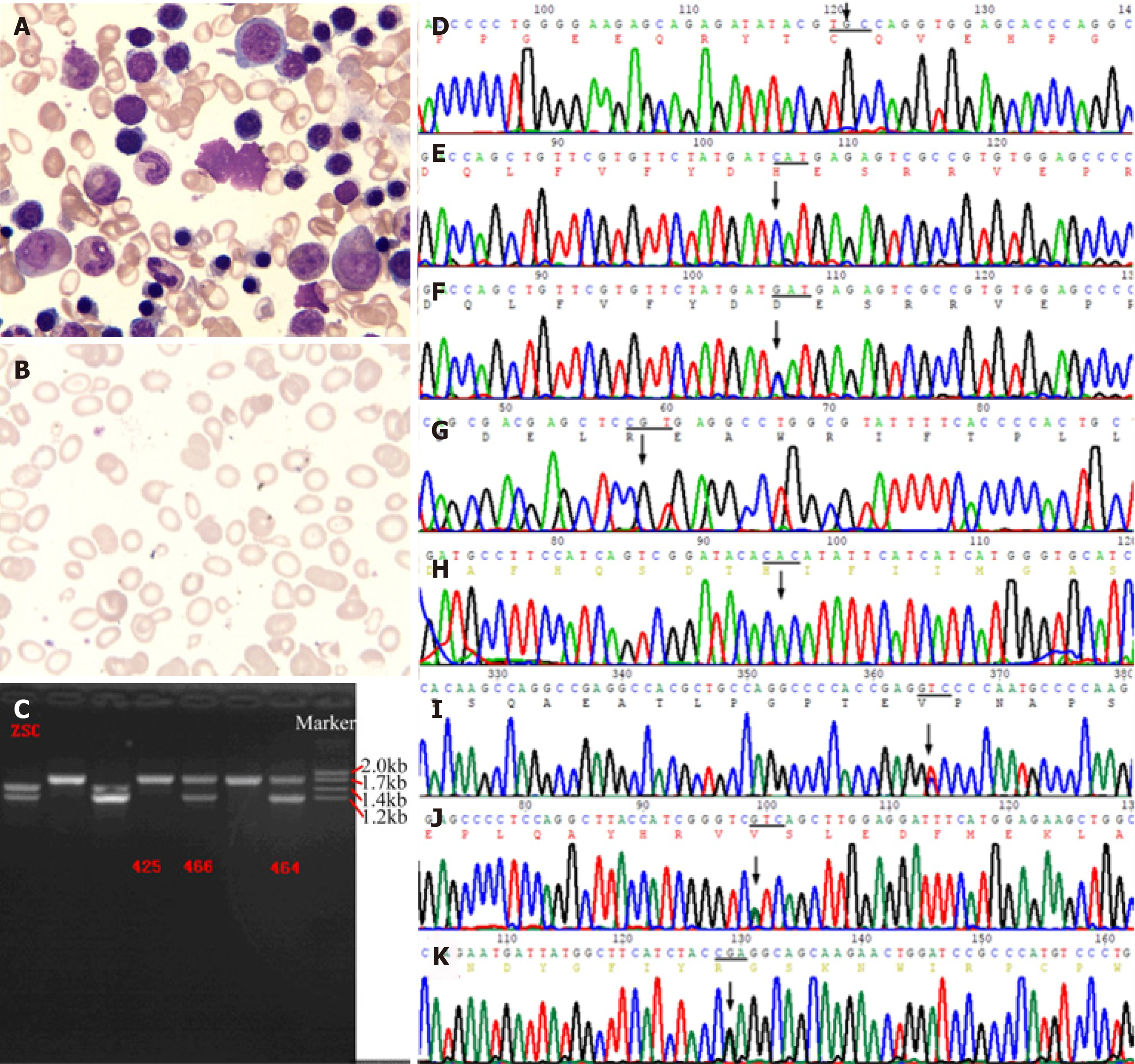
Figure 3 Bone marrow smear and genetic analysis.
A: Bone marrow smear showed erythroid cell hyperplasia, with polychromatic normoblasts. Nucleocytoplasmic development was imbalanced, with visible binuclear and mitotic types. Mature erythrocytes differed in size, which included partially large spherocytes 3%, teardrop 4%, irregular 5%, and fragmented 3% cells. Visible polychromatic erythrocytes and corpuscles were observed. Extracellular iron ++, intracellular iron -19%, +45%, ++28%, +++6%, ringed sideroblasts 2%; B: Blood smear showed obvious erythrocyte hyperplasia, possibly due to hemolytic anemia; C: Two heterozygous deletion mutants of the -α4.2, --SEA carrying globin gene were finally detected; D-H: The patient had p.C282Y and p.H63D of HFE gene, p.R459H and p.H32R of G6PD gene which were wild-type; F: His daughter carried a heterozygous p.H63D mutation of HFE gene; I-K: The patient showed p.A1583V mutation of PIEZO1 gene, p.I103V mutation of POFUT1 gene, and p.Q170R mutation of TGM5 gene.
- Citation: Ruan DD, Gan YM, Lu T, Yang X, Zhu YB, Yu QH, Liao LS, Lin N, Qian X, Luo JW, Tang FQ. Genetic diagnosis history and osteoarticular phenotype of a non-transfusion secondary hemochromatosis. World J Clin Cases 2020; 8(23): 5962-5975
- URL: https://www.wjgnet.com/2307-8960/full/v8/i23/5962.htm
- DOI: https://dx.doi.org/10.12998/wjcc.v8.i23.5962


