Copyright
©The Author(s) 2020.
World J Clin Cases. Dec 6, 2020; 8(23): 5902-5917
Published online Dec 6, 2020. doi: 10.12998/wjcc.v8.i23.5902
Published online Dec 6, 2020. doi: 10.12998/wjcc.v8.i23.5902
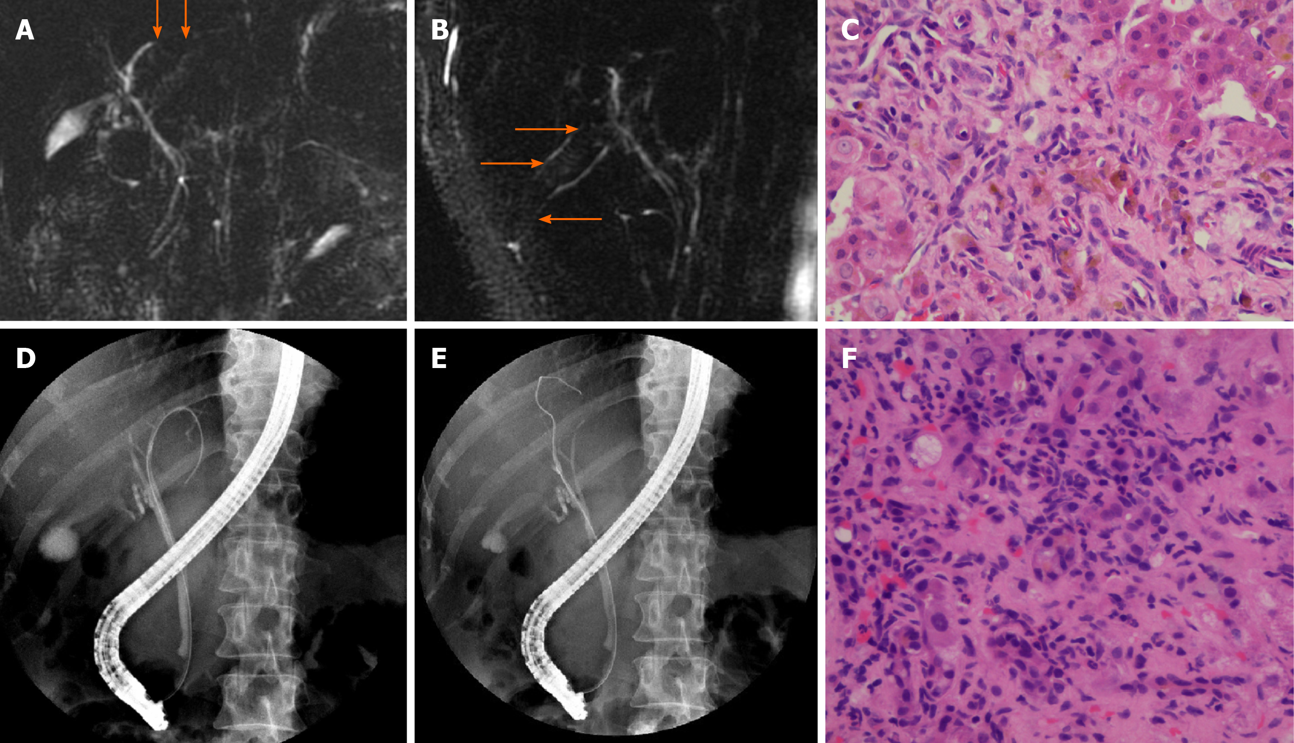
Figure 6 Magnetic resonance cholangiopancreatography images (A and B) and pathological manifestations (C) of patient No.
5 with small duct primary sclerosing cholangitis. Magnetic resonance cholangiopancreatography showed suspected multiple strictures within the biliary tree (orange arrow). Pathological examination showed preserved lobular architecture, moderate fibrosis and inflammatory processes with proliferation of ductules and feathery degeneration of hepatocytes (C). Endoscopic retrograde cholangiopancreatography (ERCP) images (D and E) and pathological manifestations (F) of patient No. 6# with AIH. ERCP showed multiple strictures of intrahepatic bile ducts in both lobes (D and E). Pathological examination showed typical chronic cholestatic hepatitis and infiltration of lymphocytes, mononuclear cells and plasma cells in the periportal area (F).
- Citation: Zhou D, Zhang B, Zhang XY, Guan WB, Wang JD, Ma F. Focal intrahepatic strictures: A proposal classification based on diagnosis-treatment experience and systemic review. World J Clin Cases 2020; 8(23): 5902-5917
- URL: https://www.wjgnet.com/2307-8960/full/v8/i23/5902.htm
- DOI: https://dx.doi.org/10.12998/wjcc.v8.i23.5902


