Copyright
©The Author(s) 2020.
World J Clin Cases. Dec 6, 2020; 8(23): 5902-5917
Published online Dec 6, 2020. doi: 10.12998/wjcc.v8.i23.5902
Published online Dec 6, 2020. doi: 10.12998/wjcc.v8.i23.5902
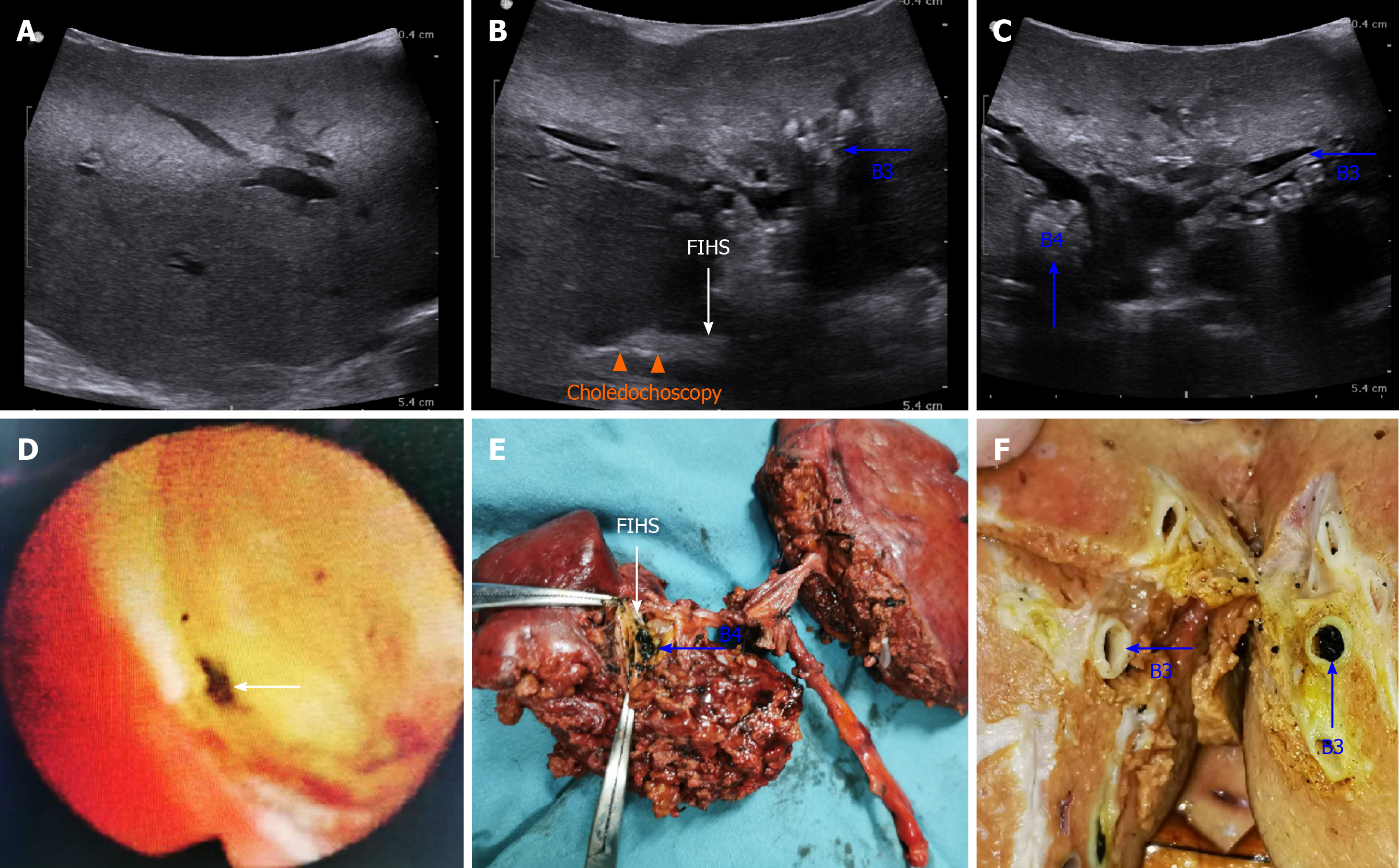
Figure 5 Intraoperative ultrasonography images of patient No.
4 (A-C). A: No dilation or stones could be detected in the intrahepatic bile ducts of the right lobe; B: Combined applications of intraoperative ultrasonography and choledochoscopy confirmed that the focal intrahepatic strictures (FIHS) was located between the peripheral part of the left hepatic duct (LHD) and the confluence of B2/B3/B4 (orange triangles). Blue arrows indicate intrahepatic stones in B3 and B4 (B and C); D: Choledochoscopy indicated FIHS at the peripheral part of the LHD; E: Specimen of the left lobe. FIHS was located at the confluence of B2/B3/B4, and stones could be seen in B4 (E) and B3 (F).
- Citation: Zhou D, Zhang B, Zhang XY, Guan WB, Wang JD, Ma F. Focal intrahepatic strictures: A proposal classification based on diagnosis-treatment experience and systemic review. World J Clin Cases 2020; 8(23): 5902-5917
- URL: https://www.wjgnet.com/2307-8960/full/v8/i23/5902.htm
- DOI: https://dx.doi.org/10.12998/wjcc.v8.i23.5902


