Copyright
©The Author(s) 2020.
World J Clin Cases. Dec 6, 2020; 8(23): 5894-5901
Published online Dec 6, 2020. doi: 10.12998/wjcc.v8.i23.5894
Published online Dec 6, 2020. doi: 10.12998/wjcc.v8.i23.5894
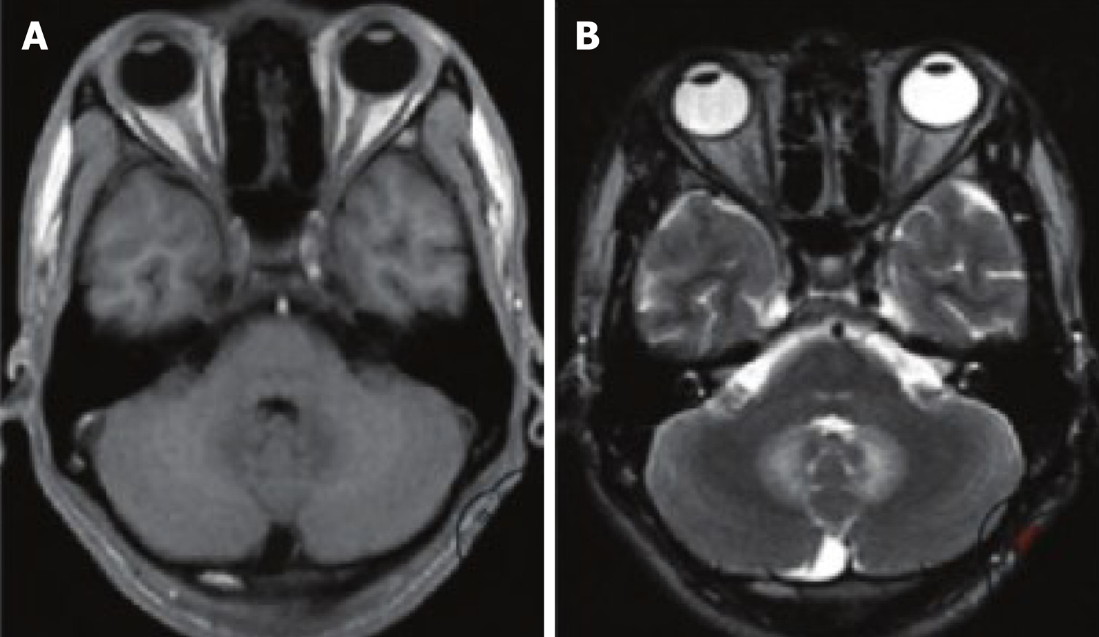
Figure 1 T1 and T2 weighted images.
A: T1 weighted image data, it can be seen that the gray matter junction of the patient's brain showed an obvious low signal shadow, the boundary is blurred and the shape is irregular; B: T2 weighted image data, an obvious high signal can be seen.
- Citation: Gu L, Yang XL, Yin HK, Lu ZH, Geng CJ. Application value analysis of magnetic resonance imaging and computed tomography in the diagnosis of intracranial infection after craniocerebral surgery. World J Clin Cases 2020; 8(23): 5894-5901
- URL: https://www.wjgnet.com/2307-8960/full/v8/i23/5894.htm
- DOI: https://dx.doi.org/10.12998/wjcc.v8.i23.5894


