Copyright
©The Author(s) 2020.
World J Clin Cases. Nov 26, 2020; 8(22): 5821-5830
Published online Nov 26, 2020. doi: 10.12998/wjcc.v8.i22.5821
Published online Nov 26, 2020. doi: 10.12998/wjcc.v8.i22.5821
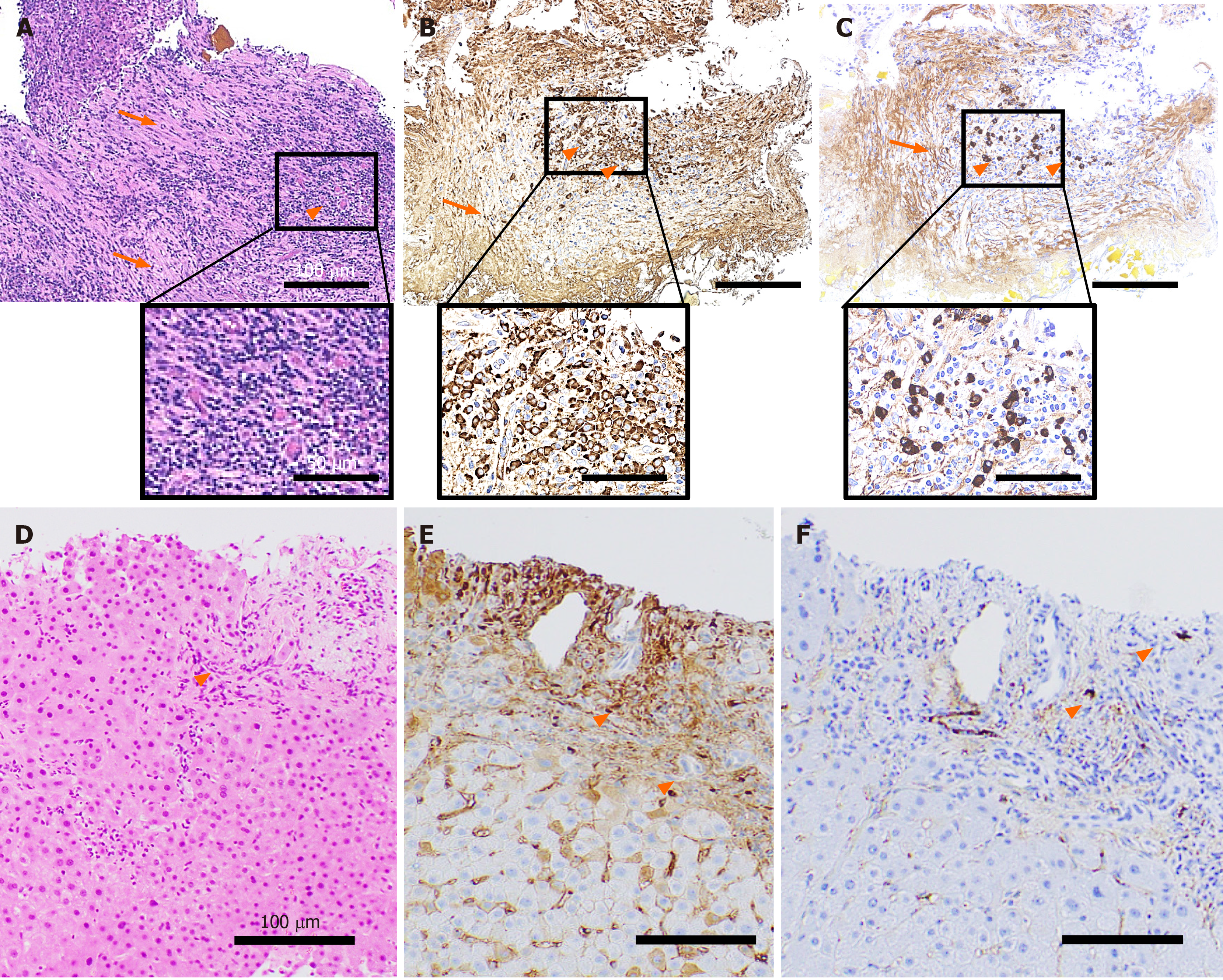
Figure 2 Histopathological findings.
A tissue sample was collected from the stenotic lower bile duct and stained with hematoxylin and eosin staining (A), IgG (B), IgG4 (C). Marked infiltration of the inflammatory cells (A-C, orange arrowheads) and storiform fibrosis (A-C, orange arrows) were observed. An increase in the number of IgG- (B) and IgG4-positive cells (C) was noted. Liver tissue showed infiltration of inflammatory cells (D: hematoxylin-eosin staining; E: IgG; F: IgG4, orange arrowheads) partly positive for IgG (E) and IgG4 (F). The scale bars represent 100 µm and 50 µm in the insets.
- Citation: Tanaka Y, Kamimura K, Nakamura R, Ohkoshi-Yamada M, Koseki Y, Mizusawa T, Ikarashi S, Hayashi K, Sato H, Sakamaki A, Yokoyama J, Terai S. Usefulness of ultrasonography to assess the response to steroidal therapy for the rare case of type 2b immunoglobulin G4-related sclerosing cholangitis without pancreatitis: A case report. World J Clin Cases 2020; 8(22): 5821-5830
- URL: https://www.wjgnet.com/2307-8960/full/v8/i22/5821.htm
- DOI: https://dx.doi.org/10.12998/wjcc.v8.i22.5821


