Copyright
©The Author(s) 2020.
World J Clin Cases. Nov 26, 2020; 8(22): 5795-5801
Published online Nov 26, 2020. doi: 10.12998/wjcc.v8.i22.5795
Published online Nov 26, 2020. doi: 10.12998/wjcc.v8.i22.5795
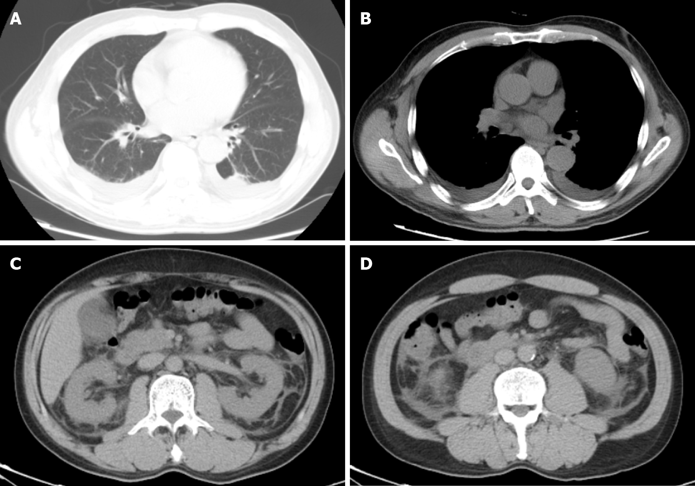
Figure 2 The computed tomography scan of chest and abdomen showed pleural effusion, perinephric effusion extended to paracolic sulcus, and slight peritoneal and pelvic effusion.
A and B: Computed tomography of the thorax and abdomen on hospital day 1 showing pleural effusion; C and D: Perinephric effusion extended to paracolic sulcus and slight peritoneal and pelvic effusion.
- Citation: Qiu FQ, Li CC, Zhou JY. Hemorrhagic fever with renal syndrome complicated with aortic dissection: A case report. World J Clin Cases 2020; 8(22): 5795-5801
- URL: https://www.wjgnet.com/2307-8960/full/v8/i22/5795.htm
- DOI: https://dx.doi.org/10.12998/wjcc.v8.i22.5795


