Copyright
©The Author(s) 2020.
World J Clin Cases. Nov 26, 2020; 8(22): 5781-5789
Published online Nov 26, 2020. doi: 10.12998/wjcc.v8.i22.5781
Published online Nov 26, 2020. doi: 10.12998/wjcc.v8.i22.5781
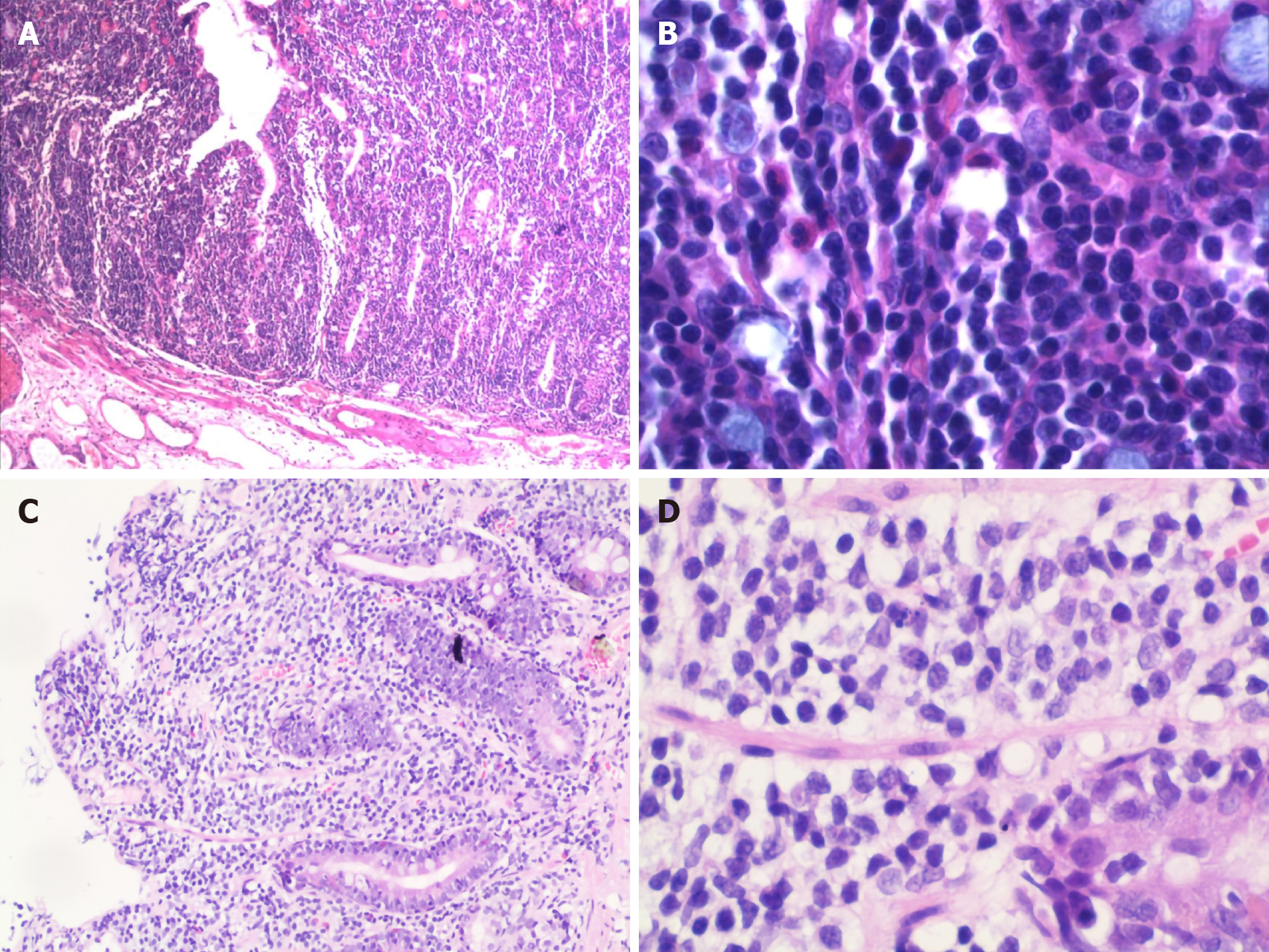
Figure 4 Pathology of the colon and duodenum A: Descending colon.
Villous adenoma with focal carcinogenesis (moderately differentiated adenocarcinoma), limited to the mucosal layer, was observed (× 100); B: Descending colon. There was diffuse atypical lymphocyte proliferation with a single form mainly infiltrated in the mucosal epithelium and lamina propria, partly invading the submucosa (× 400); C: Duodenum. Several lymphocytes infiltrated in the mucosal layer (× 100); D: Duodenum. Atypical lymphocytes accumulated in the mucosal layer with medium-sized cell bodies consisting of few cytoplasm and large oval or irregular nucleus containing coarse chromatin (× 400).
- Citation: Zhang MY, Min CC, Fu WW, Liu H, Yin XY, Zhang CP, Tian ZB, Li XY. Early colon cancer with enteropathy-associated T-cell lymphoma involving the whole gastrointestinal tract: A case report. World J Clin Cases 2020; 8(22): 5781-5789
- URL: https://www.wjgnet.com/2307-8960/full/v8/i22/5781.htm
- DOI: https://dx.doi.org/10.12998/wjcc.v8.i22.5781


