Copyright
©The Author(s) 2020.
World J Clin Cases. Nov 26, 2020; 8(22): 5781-5789
Published online Nov 26, 2020. doi: 10.12998/wjcc.v8.i22.5781
Published online Nov 26, 2020. doi: 10.12998/wjcc.v8.i22.5781
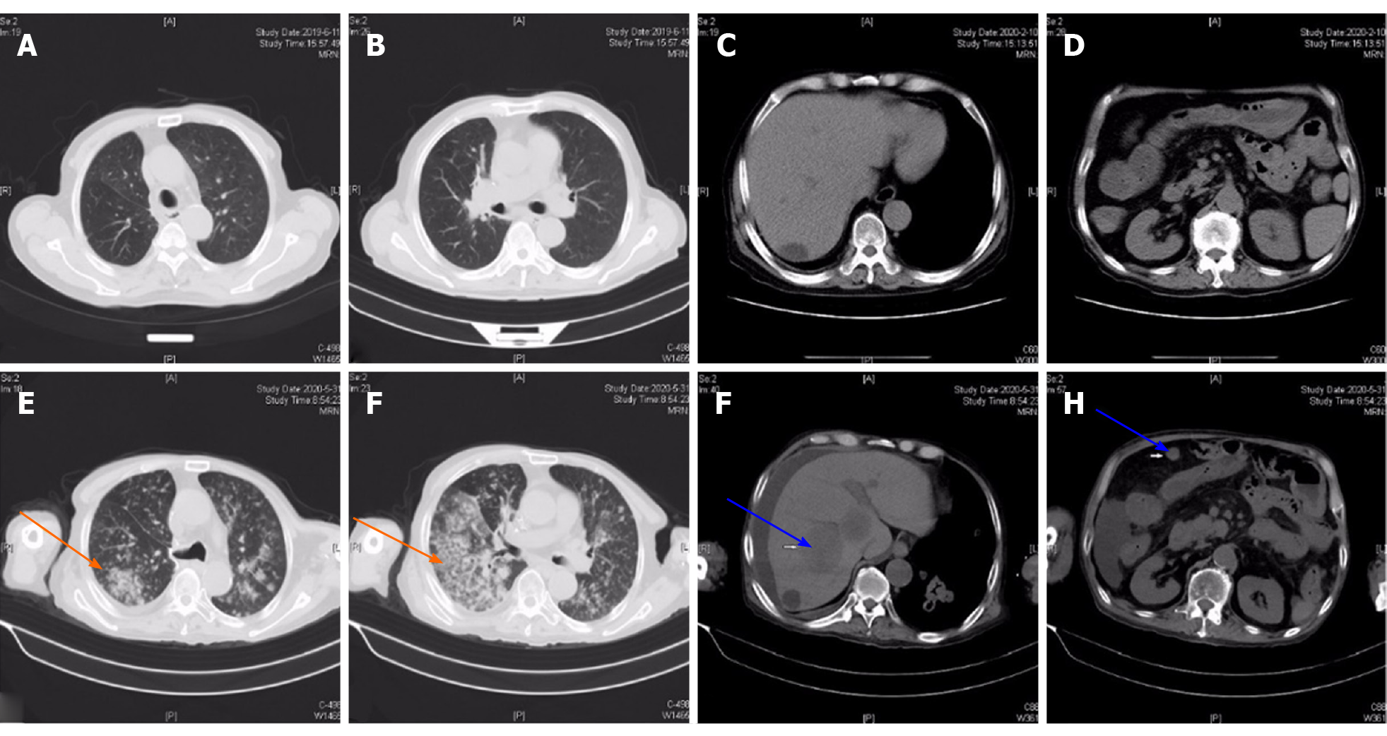
Figure 1 Chest and abdominal computed tomography showed rapid disease progression.
A and B: Chest computed tomography (CT) on June 11, 2019 showed clear lung fields on both sides; C and D: Abdominal CT on June 11, 2019 showed a liver cyst, and part of a slightly dilated small intestine; E and F: Chest CT on May 31, 2020 revealed multiple nodules and masses in both lungs, not excluding neoplastic lesions (orange arrow); G and H: Abdominal CT on May 31, 2020 showed multiple low-density lesions in the right liver and multiple soft tissue masses in the abdominal cavity, suggesting multiple metastases (blue arrow).
- Citation: Zhang MY, Min CC, Fu WW, Liu H, Yin XY, Zhang CP, Tian ZB, Li XY. Early colon cancer with enteropathy-associated T-cell lymphoma involving the whole gastrointestinal tract: A case report. World J Clin Cases 2020; 8(22): 5781-5789
- URL: https://www.wjgnet.com/2307-8960/full/v8/i22/5781.htm
- DOI: https://dx.doi.org/10.12998/wjcc.v8.i22.5781


