Copyright
©The Author(s) 2020.
World J Clin Cases. Nov 26, 2020; 8(22): 5729-5736
Published online Nov 26, 2020. doi: 10.12998/wjcc.v8.i22.5729
Published online Nov 26, 2020. doi: 10.12998/wjcc.v8.i22.5729
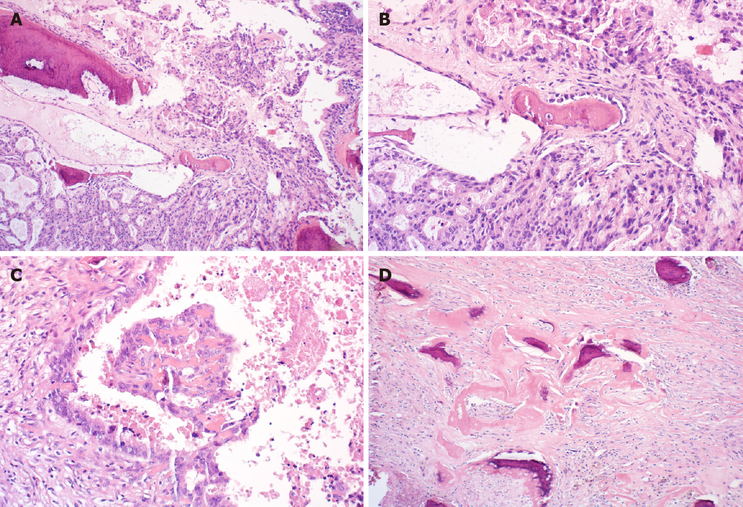
Figure 2 Postoperative pathological examination.
A: Atypical epithelial cribriform hyperplasia was seen in some areas [hematoxylin and eosin (HE) 100 ×]; B: Protrusion into the cystic cavity with massive necrosis and hemorrhage; multifocal pericancerous interstitial ossification and infiltration into the fibrous stroma (HE 200 ×); C: Several foci of cancer cells produced a bone-like matrix (HE 200 ×); D: Part of the cyst wall thickening and fibrosis with previous hemorrhage and hemosiderin deposition and interstitial mass of fibrous bone (HE 100 ×).
- Citation: Gao KJ, Yan ZL, Yu Y, Guo LQ, Hang C, Yang JB, Zhang MC. Port-site metastasis of unsuspected gallbladder carcinoma with ossification after laparoscopic cholecystectomy: A case report. World J Clin Cases 2020; 8(22): 5729-5736
- URL: https://www.wjgnet.com/2307-8960/full/v8/i22/5729.htm
- DOI: https://dx.doi.org/10.12998/wjcc.v8.i22.5729


