Copyright
©The Author(s) 2020.
World J Clin Cases. Nov 26, 2020; 8(22): 5690-5700
Published online Nov 26, 2020. doi: 10.12998/wjcc.v8.i22.5690
Published online Nov 26, 2020. doi: 10.12998/wjcc.v8.i22.5690
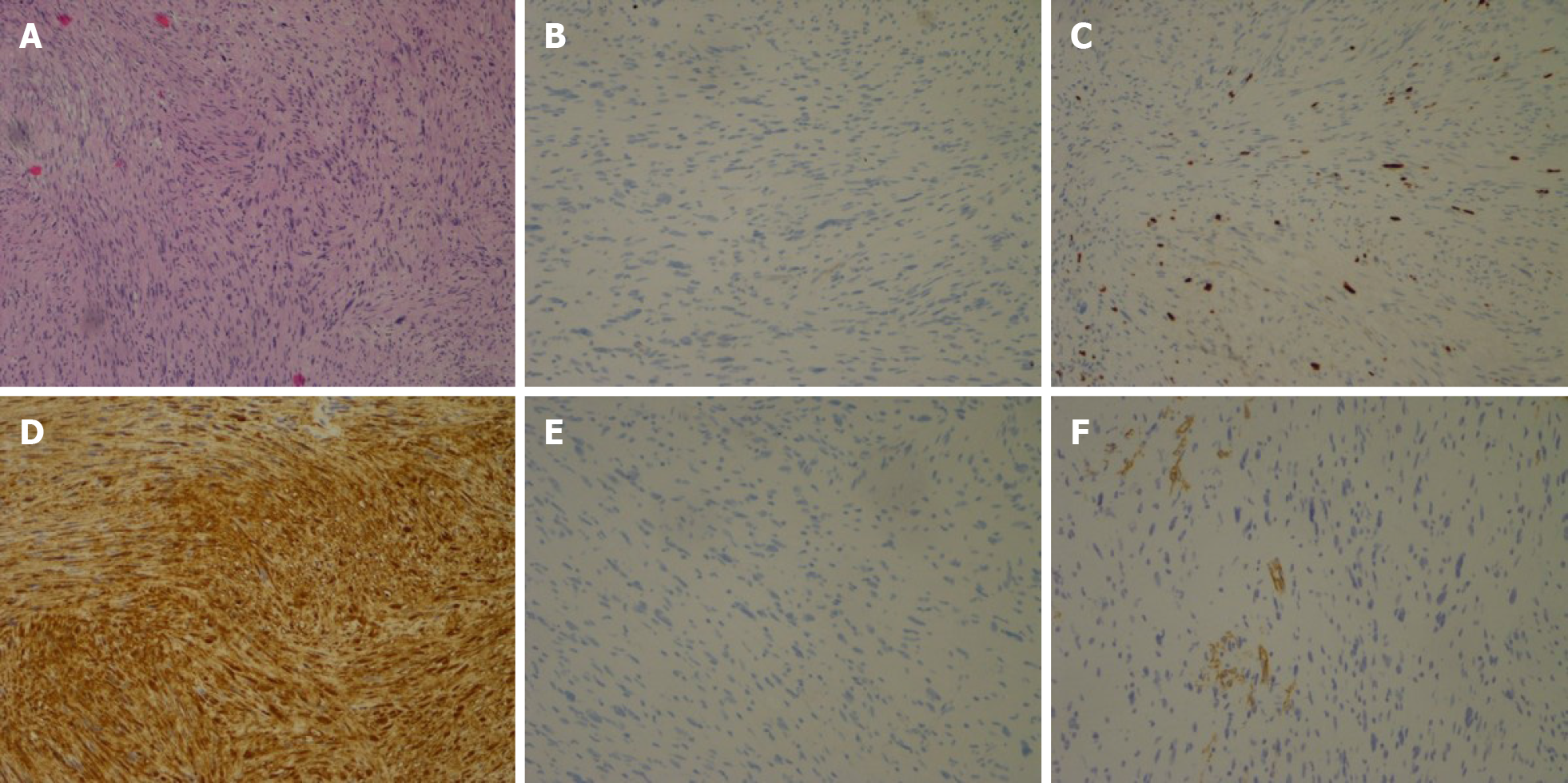
Figure 5 Endoscopic submucosal excision performed.
A: Histopathological examination of the tumor in case 1 showed spindle-shaped cells arranged in bundles; B: Immunochemical analysis revealed no staining with CD117; C: Focal positivity with CD34; D: Positive staining with S100; E: DOG-1; F: The mitotic activity was 3 mitosis/50 high-power field on Ki-67 staining.
- Citation: Li B, Wang X, Zou WL, Yu SX, Chen Y, Xu HW. Endoscopic resection of benign esophageal schwannoma: Three case reports and review of literature. World J Clin Cases 2020; 8(22): 5690-5700
- URL: https://www.wjgnet.com/2307-8960/full/v8/i22/5690.htm
- DOI: https://dx.doi.org/10.12998/wjcc.v8.i22.5690


