Copyright
©The Author(s) 2020.
World J Clin Cases. Nov 26, 2020; 8(22): 5684-5689
Published online Nov 26, 2020. doi: 10.12998/wjcc.v8.i22.5684
Published online Nov 26, 2020. doi: 10.12998/wjcc.v8.i22.5684
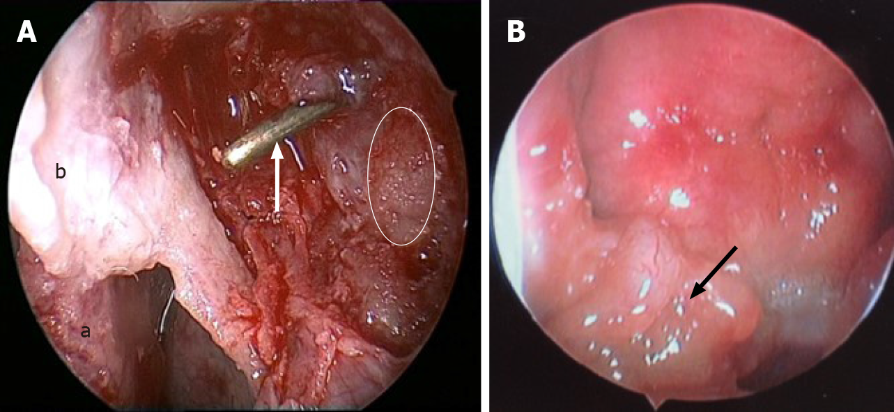
Figure 4 Endoscopic images in surgery.
A: The left lacrimal sac was filled with friable, grey to yellow, glistening, and waxy sediment (white line area), dacryocystorhinostomy was done, and the probe (white arrow) showed opening of the lacrimal sac into the nasal cavity (a: Middle turbinate; b: Nasal mucosa flap); B: An irregular-surfaced mass was noted in the right nasopharynx.
- Citation: Song X, Yang J, Lai Y, Zhou J, Wang J, Sun X, Wang D. Localized amyloidosis affecting the lacrimal sac managed by endoscopic surgery: A case report. World J Clin Cases 2020; 8(22): 5684-5689
- URL: https://www.wjgnet.com/2307-8960/full/v8/i22/5684.htm
- DOI: https://dx.doi.org/10.12998/wjcc.v8.i22.5684


