Copyright
©The Author(s) 2020.
World J Clin Cases. Nov 26, 2020; 8(22): 5684-5689
Published online Nov 26, 2020. doi: 10.12998/wjcc.v8.i22.5684
Published online Nov 26, 2020. doi: 10.12998/wjcc.v8.i22.5684
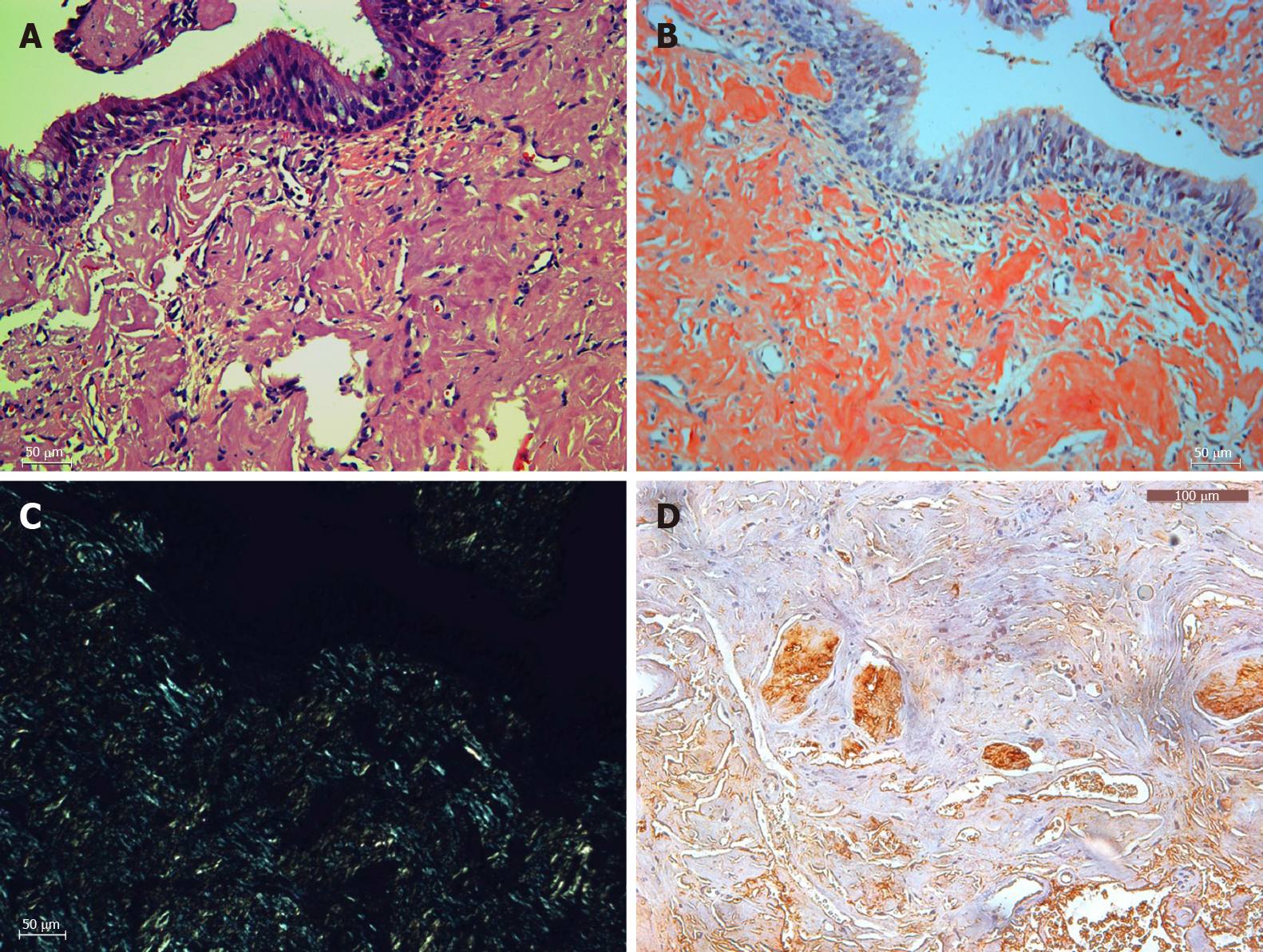
Figure 3 Histopathology (× 200).
A: Hematoxylin and eosin staining of amyloid tissues; B: Congo staining of the same area of amyloid tissues; C: Corresponding area under a polarization microscope; D: Immunohistochemistry showed positive lambda light chain staining. Bar: 100 μm.
- Citation: Song X, Yang J, Lai Y, Zhou J, Wang J, Sun X, Wang D. Localized amyloidosis affecting the lacrimal sac managed by endoscopic surgery: A case report. World J Clin Cases 2020; 8(22): 5684-5689
- URL: https://www.wjgnet.com/2307-8960/full/v8/i22/5684.htm
- DOI: https://dx.doi.org/10.12998/wjcc.v8.i22.5684


