Copyright
©The Author(s) 2020.
World J Clin Cases. Nov 26, 2020; 8(22): 5684-5689
Published online Nov 26, 2020. doi: 10.12998/wjcc.v8.i22.5684
Published online Nov 26, 2020. doi: 10.12998/wjcc.v8.i22.5684
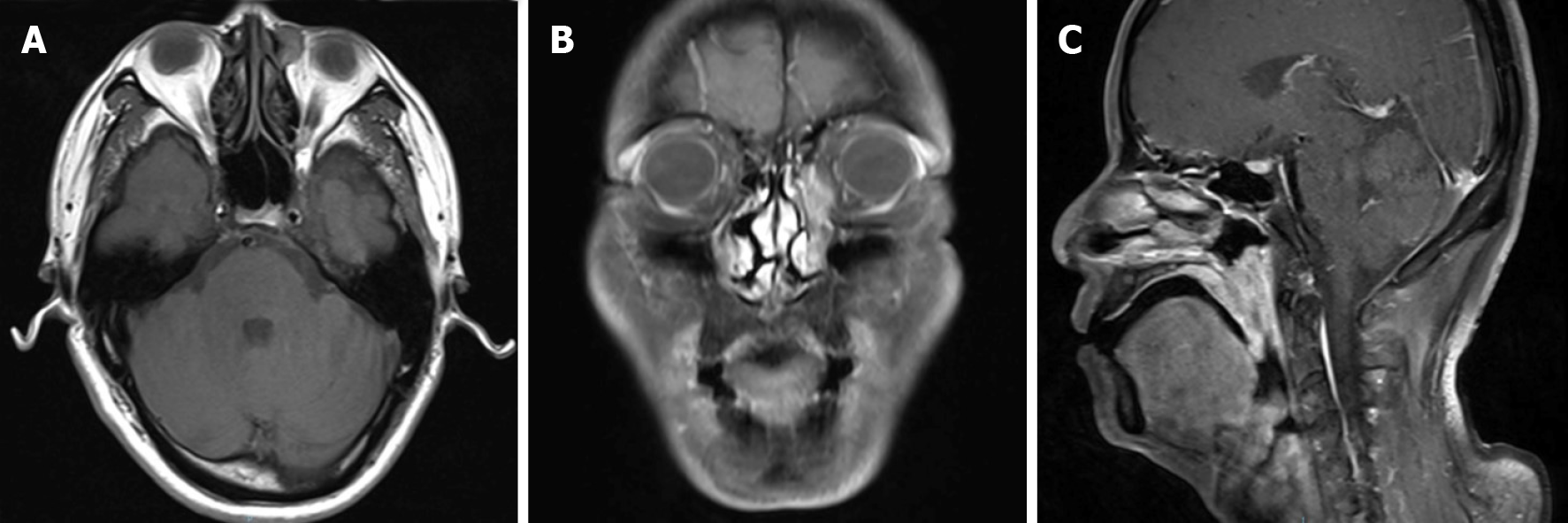
Figure 2 Pre-operative head and neck magnetic resonance imaging.
A: T1 weighted imaging (T1WI) showed a round soft tissue intensity mass within the left lacrimal sac, presenting equal signal in T1WI, and heterogeneous high signal in T2WI, without obvious enhancement when contrasted; B: T1WI with contrast showed an abnormal high signal mass along the left nasolacrimal duct, from the lacrimal sac to the level of inferior turbinate; C: Sagittal T1WI demonstrated extensive soft tissue thickening of the posterior wall of the nasopharynx and the lateral wall of the oropharynx and uvula, with isointensity on T1WI and slightly high signal on T2WI, apparently enhanced when contrasted.
- Citation: Song X, Yang J, Lai Y, Zhou J, Wang J, Sun X, Wang D. Localized amyloidosis affecting the lacrimal sac managed by endoscopic surgery: A case report. World J Clin Cases 2020; 8(22): 5684-5689
- URL: https://www.wjgnet.com/2307-8960/full/v8/i22/5684.htm
- DOI: https://dx.doi.org/10.12998/wjcc.v8.i22.5684


