Copyright
©The Author(s) 2020.
World J Clin Cases. Nov 26, 2020; 8(22): 5645-5656
Published online Nov 26, 2020. doi: 10.12998/wjcc.v8.i22.5645
Published online Nov 26, 2020. doi: 10.12998/wjcc.v8.i22.5645
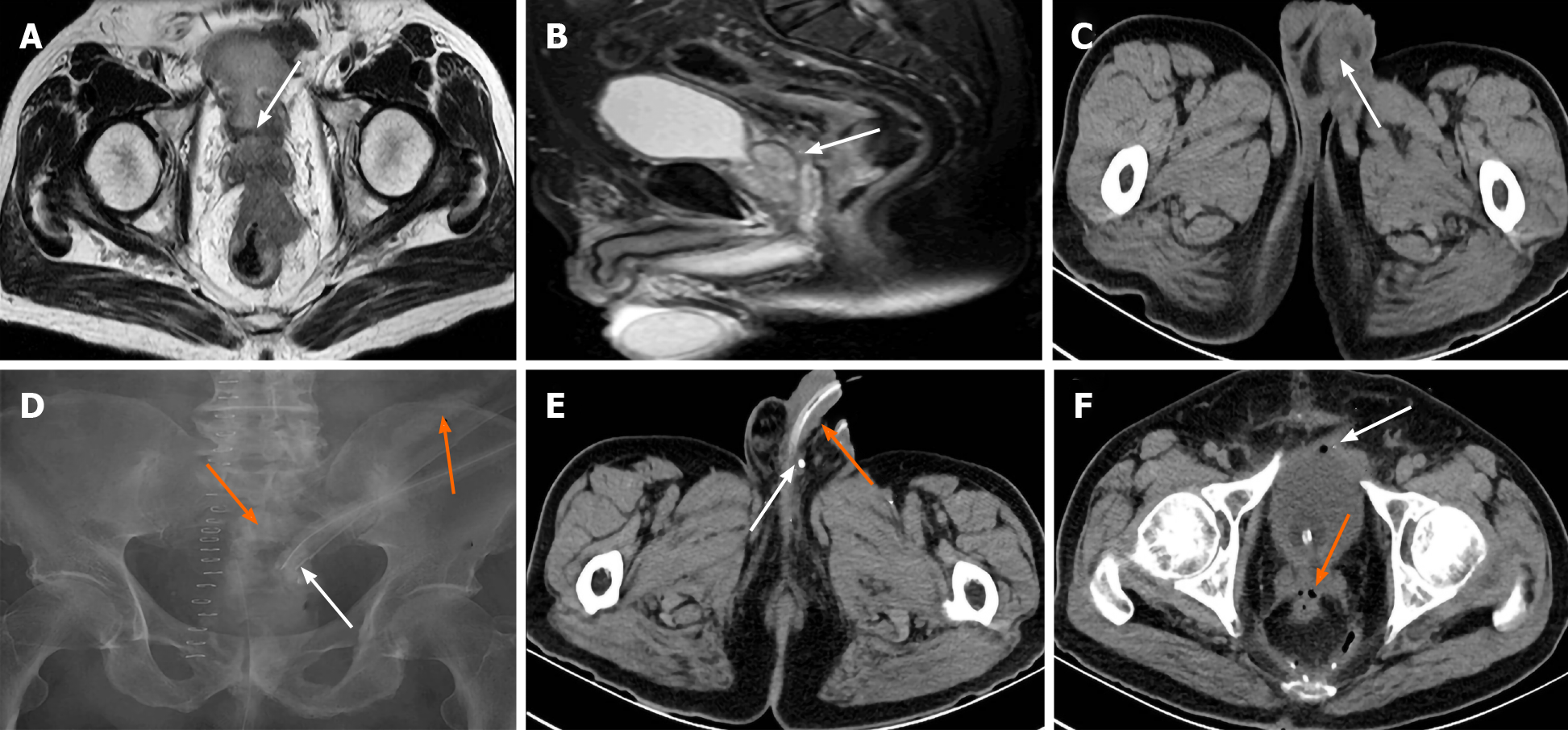
Figure 4 Imaging findings of case 3.
A, B: Cross-section and sagittal plane magnetic resonance imaging (MRI) showed that the bladder was filled well, coating of the seminal vesicles (SV) were complete, the huge rectal tumor invaded Denonvilliers’ fascia (white arrows) at the level of the SVs; C: The left swollen and infective scrotum (white arrow) by computed tomography (CT); D: Transabdominal sinus radiography showed contrast agent entering the rectum and proximal colon (orange arrows) through intrapelvic drainage tube (white arrow); E: Contrast agent residue (white arrow) in the ductus deferens beside the entrance to the epididymis but not urethra (orange arrow) by CT; F: A few effusion and bubbles in the pelvic cavity, some bubbles entering the SV (orange arrow) and bladder (white arrow) via the sinus from rectal anastomotic leakage.
- Citation: Xia ZX, Cong JC, Zhang H. Rectoseminal vesicle fistula after radical surgery for rectal cancer: Four case reports and a literature review. World J Clin Cases 2020; 8(22): 5645-5656
- URL: https://www.wjgnet.com/2307-8960/full/v8/i22/5645.htm
- DOI: https://dx.doi.org/10.12998/wjcc.v8.i22.5645


