Copyright
©The Author(s) 2020.
World J Clin Cases. Nov 26, 2020; 8(22): 5645-5656
Published online Nov 26, 2020. doi: 10.12998/wjcc.v8.i22.5645
Published online Nov 26, 2020. doi: 10.12998/wjcc.v8.i22.5645
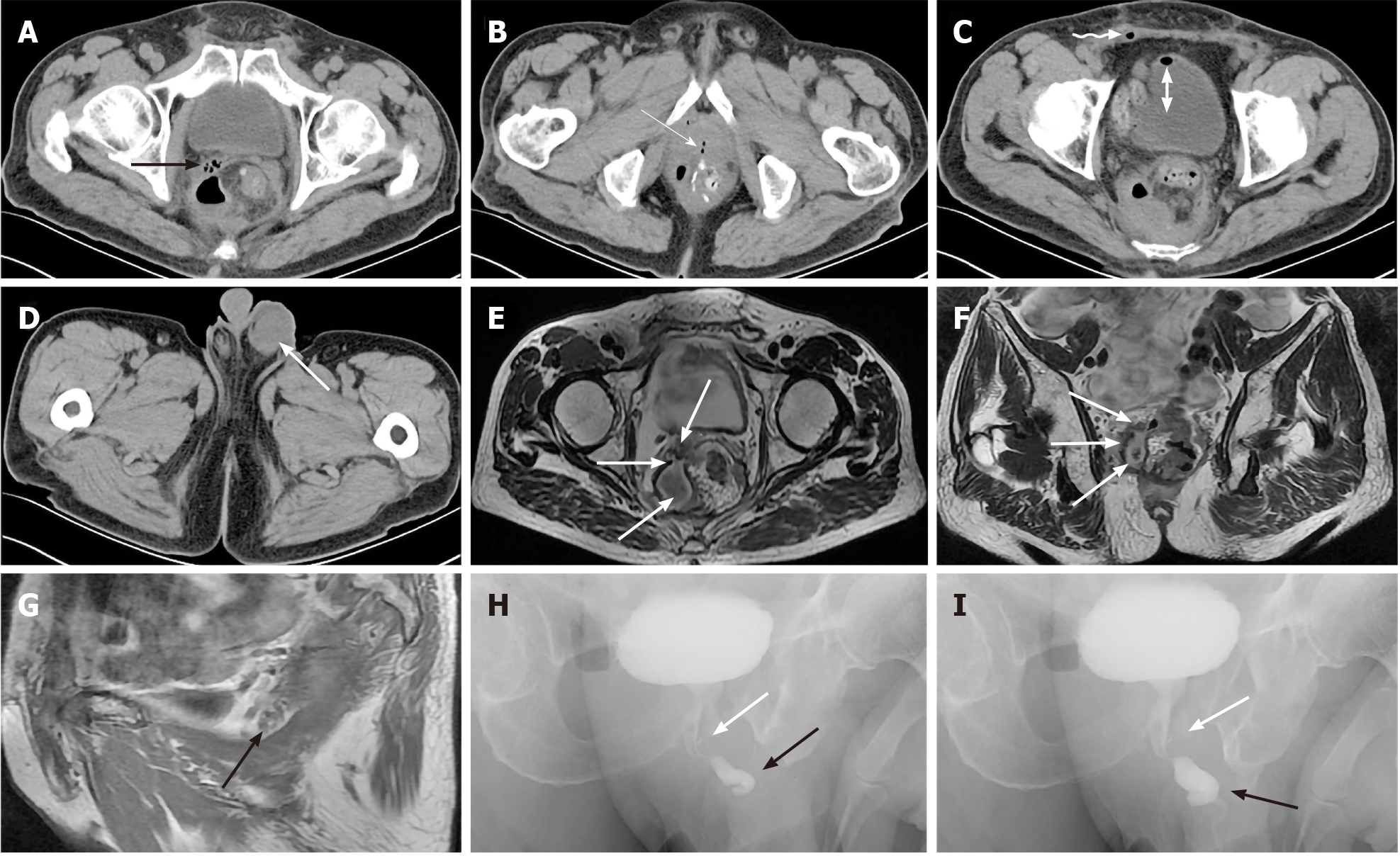
Figure 3 Imaging findings of case 2.
A-C: Computed tomography demonstrates an encapsulated cavity filled with air bubbles, pus and effusion with an air-water level connecting with the right seminal vesicles (SV) (black arrow) and ejaculatory duct opening of the urethral prostate caruncle (white arrow) after anastomotic leakage (AL). Some bubbles in the right deferent duct within the spermatic cord (curved arrow) and some in the bladder (double arrow); D: Left edematous and infective scrotum (white arrow); E, F: Cross-section and coronal plane magnetic resonance imaging (MRI) displayed a sinus between encapsulated pus cavity and right edematous SV (white arrows) secondary to AL; G: Sagittal plane MRI demonstrated some bubbles in the right edematous SV (black arrow) between bladder and rectum; H, I: Urethral retrograde radiography showed that the bladder was well filled, the anterior urethra (black arrows) was normal, the posterior urethra was slightly narrow, and the ejaculatory duct was filled with a small amount of contrast agent (white arrows).
- Citation: Xia ZX, Cong JC, Zhang H. Rectoseminal vesicle fistula after radical surgery for rectal cancer: Four case reports and a literature review. World J Clin Cases 2020; 8(22): 5645-5656
- URL: https://www.wjgnet.com/2307-8960/full/v8/i22/5645.htm
- DOI: https://dx.doi.org/10.12998/wjcc.v8.i22.5645


