Copyright
©The Author(s) 2020.
World J Clin Cases. Nov 26, 2020; 8(22): 5645-5656
Published online Nov 26, 2020. doi: 10.12998/wjcc.v8.i22.5645
Published online Nov 26, 2020. doi: 10.12998/wjcc.v8.i22.5645
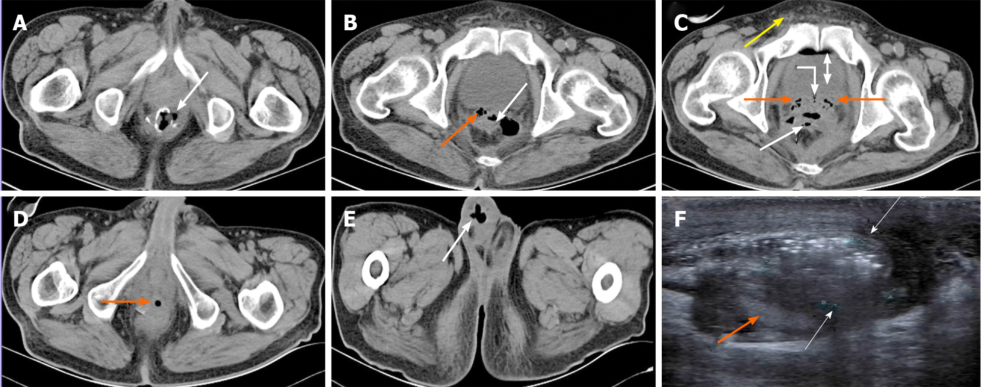
Figure 2 Imaging findings of case 1.
A-C: X-ray computed tomography (CT) demonstrated edematous rectal wall and double seminal vesicles (SV), incomplete anastomosis with an obvious rectal anastomotic leakage (AL) (white arrows). A large pelvic cavity around the AL formed a sinus from the rectum to SV, and air bubbles entered bilateral SVs (orange arrows), and ampulla of deferent duct (curved arrow). Some bubbles present in the bladder (double arrow)and a few in the right deferent duct within the spermatic cord (yellow arrow); D: CT displayed air bubbles (orange arrow) located within ejaculatory duct opening of the urethral prostate; E: Bubbles (white arrow) from the right sperm duct retrogradely entered the right edematous and infected scrotum; F: Urinary system color Doppler ultrasound showed a right epididymis tail 2.2 cm x 1.8 cm x 1.5 cm enclosed abscess and another small abscess in the right edematous scrotum with a blurred boundary, a number of strong gas echoes and peripheral rich blood (white arrows) around the testis and epididymis (orange arrow).
- Citation: Xia ZX, Cong JC, Zhang H. Rectoseminal vesicle fistula after radical surgery for rectal cancer: Four case reports and a literature review. World J Clin Cases 2020; 8(22): 5645-5656
- URL: https://www.wjgnet.com/2307-8960/full/v8/i22/5645.htm
- DOI: https://dx.doi.org/10.12998/wjcc.v8.i22.5645


