Copyright
©The Author(s) 2020.
World J Clin Cases. Nov 26, 2020; 8(22): 5632-5638
Published online Nov 26, 2020. doi: 10.12998/wjcc.v8.i22.5632
Published online Nov 26, 2020. doi: 10.12998/wjcc.v8.i22.5632
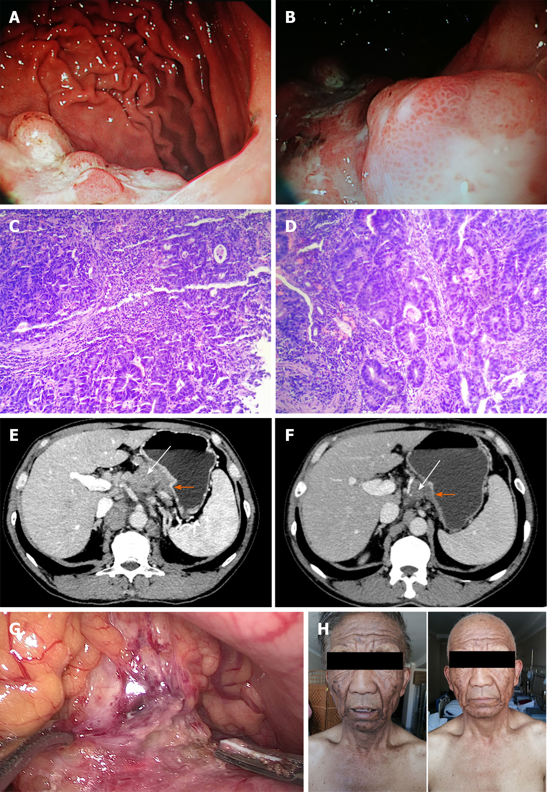
Figure 2 Patient’s examination results and skin changes before and after treatment.
A and B: Gastroscopy at Xijing Hospital: Gastroscopy of the upper part of the stomach under the patient’s cardia showed a large mucous membrane bulge near the posterior wall, with an uneven surface and large areas of ulceration. The biopsy was brittle, and a diagnosis of cardiac gastric cancer was made; C and D: A gastroscopic biopsy at Xijing Hospital showed that the heteromorphic cells had been arranged in glandular, ribbon-like, and lamellar patterns, with obvious atypia and invasive growth. Diagnosis: Cancer of the gastric cardia with medium-poorly differentiated adenocarcinoma (C: Areas of poorly differentiated adenocarcinoma, hematoxylin–eosin staining: 200 ×; D: Areas of medium differentiated adenocarcinoma, hematoxylin–eosin staining: 200 ×); E: Computed tomography scan before neoadjuvant chemotherapy showed thickening of the gastric cardia wall (orange arrow) and enlargement of the peripheral lymph nodes (white arrow); F: After three cycles of neoadjuvant chemotherapy, computed tomography scan showed that the tumor (orange arrow) and surrounding lymph nodes (white arrow) were significantly reduced; G: Tumor invasion of the pancreas was observed during surgery; H: After three cycles of chemotherapy, skin pigmentation was reduced.
- Citation: Wang N, Yu PJ, Liu ZL, Zhu SM, Zhang CW. Malignant acanthosis nigricans with Leser–Trélat sign and tripe palms: A case report. World J Clin Cases 2020; 8(22): 5632-5638
- URL: https://www.wjgnet.com/2307-8960/full/v8/i22/5632.htm
- DOI: https://dx.doi.org/10.12998/wjcc.v8.i22.5632


