Copyright
©The Author(s) 2020.
World J Clin Cases. Nov 26, 2020; 8(22): 5632-5638
Published online Nov 26, 2020. doi: 10.12998/wjcc.v8.i22.5632
Published online Nov 26, 2020. doi: 10.12998/wjcc.v8.i22.5632
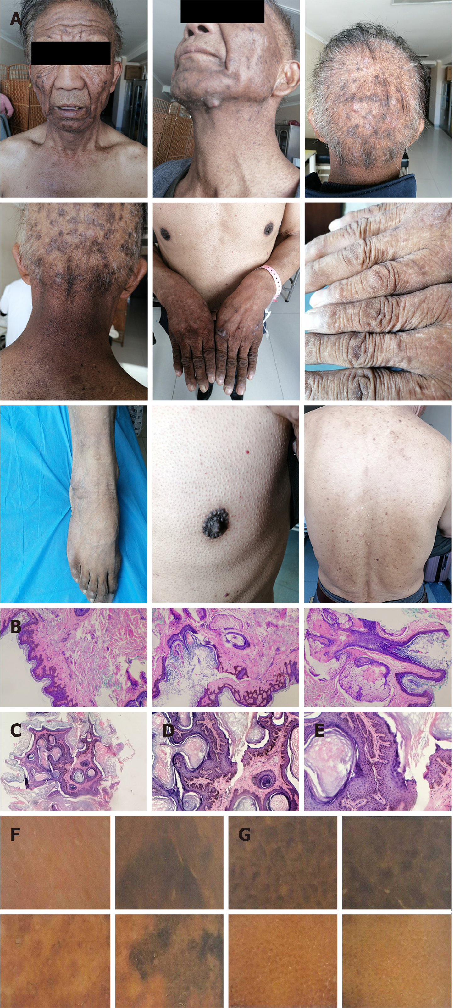
Figure 1 Patient’s skin appearance and examination results.
A: Skin lesions; B: Cervical pathology: Epidermal hyperkeratosis, basal cell hyperplasia, pigmentation, and papilla protrusion (hematoxylin–eosin staining: 100 ×); C-E: Facial flat brown maculopapular pathology: Basal cell papilloma, hyperpigmentation, and hyperkeratosis (C: Hematoxylin–eosin staining: 40 ×; D: Hematoxylin–eosin staining: 100 ×; E: Hematoxylin–eosin staining: 200 ×); F and G: A dermoscopic examination of the surface of the hand showed consistent gray–brown papillary hyperplasia and diffuse dark brown to black–gray pigmentation with slight scales.
- Citation: Wang N, Yu PJ, Liu ZL, Zhu SM, Zhang CW. Malignant acanthosis nigricans with Leser–Trélat sign and tripe palms: A case report. World J Clin Cases 2020; 8(22): 5632-5638
- URL: https://www.wjgnet.com/2307-8960/full/v8/i22/5632.htm
- DOI: https://dx.doi.org/10.12998/wjcc.v8.i22.5632


