Copyright
©The Author(s) 2020.
World J Clin Cases. Nov 26, 2020; 8(22): 5555-5563
Published online Nov 26, 2020. doi: 10.12998/wjcc.v8.i22.5555
Published online Nov 26, 2020. doi: 10.12998/wjcc.v8.i22.5555
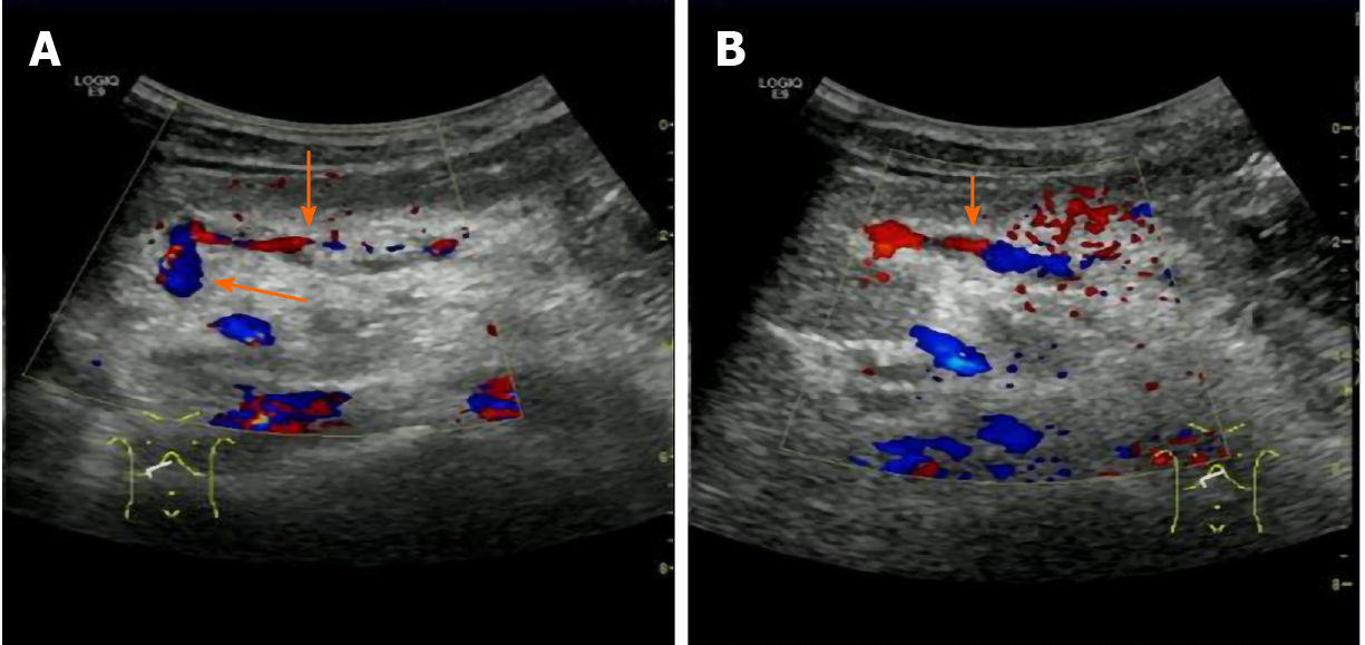
Figure 1 Color Doppler ultrasonography of a 9-year-old boy after recanalized umbilical vein as a conduit for Rex shunt (gastric coronary vein-umbilical vein shunt).
A: Color Doppler flow imaging (CDFI)showed intermittent blood flow signal in the bypass vessel (gastric coronary vein) 7 d after operation; B: CDFI showed that the bypass vessel was well filled with blood flow signals after clinical anticoagulation treatment for 3 mo. Long arrow indicates umbilical vein and short arrow indicates gastric coronary vein.
- Citation: Zhang YQ, Wang Q, Wu M, Li Y, Wei XL, Zhang FX, Li Y, Shao GR, Xiao J. Sonographic features of umbilical vein recanalization for a Rex shunt on cavernous transformation of portal vein in children . World J Clin Cases 2020; 8(22): 5555-5563
- URL: https://www.wjgnet.com/2307-8960/full/v8/i22/5555.htm
- DOI: https://dx.doi.org/10.12998/wjcc.v8.i22.5555


