Copyright
©The Author(s) 2020.
World J Clin Cases. Nov 26, 2020; 8(22): 5535-5546
Published online Nov 26, 2020. doi: 10.12998/wjcc.v8.i22.5535
Published online Nov 26, 2020. doi: 10.12998/wjcc.v8.i22.5535
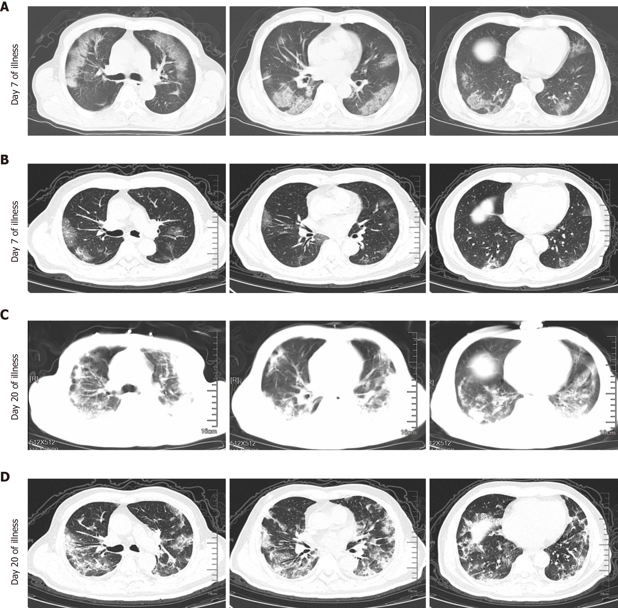
Figure 1 Chest computed tomography of critical coronavirus disease 2019 patients with different severity.
A: Chest computed tomography (CT) of a 71-year-old man (non-survivor, case 1) showed multifocal and bilateral ground-glass opacities (GGO) in the alveolitis stage (Day 7 of illness); B: Chest CT of a 73-year-old male patient (survivor, case 2) exhibited slight GGO in the alveolitis stage (Day 7 of illness); C: Classified into the fibrosis stage (Day 20 of illness) and Chest CT (case 1) showed bilateral massive shadows of high density and GGO, accompanied by the air bronchogram sign and reticular pattern in the fibrosis stage; and D: Chest CT (case 2) showed that bilateral and multifocal lesions were observed with a combination of mixed GGO, reticular pattern, bronchiectasis and few consolidation (Day 20 of illness).
- Citation: Lv XT, Zhu YP, Cheng AG, Jin YX, Ding HB, Wang CY, Zhang SY, Chen GP, Chen QQ, Liu QC. High serum lactate dehydrogenase and dyspnea: Positive predictors of adverse outcome in critical COVID-19 patients in Yichang. World J Clin Cases 2020; 8(22): 5535-5546
- URL: https://www.wjgnet.com/2307-8960/full/v8/i22/5535.htm
- DOI: https://dx.doi.org/10.12998/wjcc.v8.i22.5535


