Copyright
©The Author(s) 2020.
World J Clin Cases. Nov 26, 2020; 8(22): 5501-5512
Published online Nov 26, 2020. doi: 10.12998/wjcc.v8.i22.5501
Published online Nov 26, 2020. doi: 10.12998/wjcc.v8.i22.5501
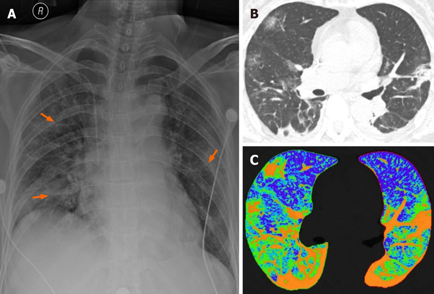
Figure 1 Chest radiography and computed tomography images of a 65-year-old coronavirus disease-2019 patient.
A: Posteroanterior chest radiography showed multiple patchy high density shadows (orange arrow) in the outer field of bilateral lungs; B: The thin-section transverse computed tomography image revealed multiple ground-glass opacities, consolidation, fibrosis and thickened pleura in the peripheral area of both lungs; C: The two-dimensional pseudocolor reconstruction image highlighted the distribution and range of pulmonary lesions (orange area).
- Citation: Tang L, Wang Y, Zhang Y, Zhang XY, Zeng XC, Song B. COVID-19: A review of what radiologists need to know. World J Clin Cases 2020; 8(22): 5501-5512
- URL: https://www.wjgnet.com/2307-8960/full/v8/i22/5501.htm
- DOI: https://dx.doi.org/10.12998/wjcc.v8.i22.5501


