Copyright
©The Author(s) 2020.
World J Clin Cases. Nov 6, 2020; 8(21): 5313-5319
Published online Nov 6, 2020. doi: 10.12998/wjcc.v8.i21.5313
Published online Nov 6, 2020. doi: 10.12998/wjcc.v8.i21.5313
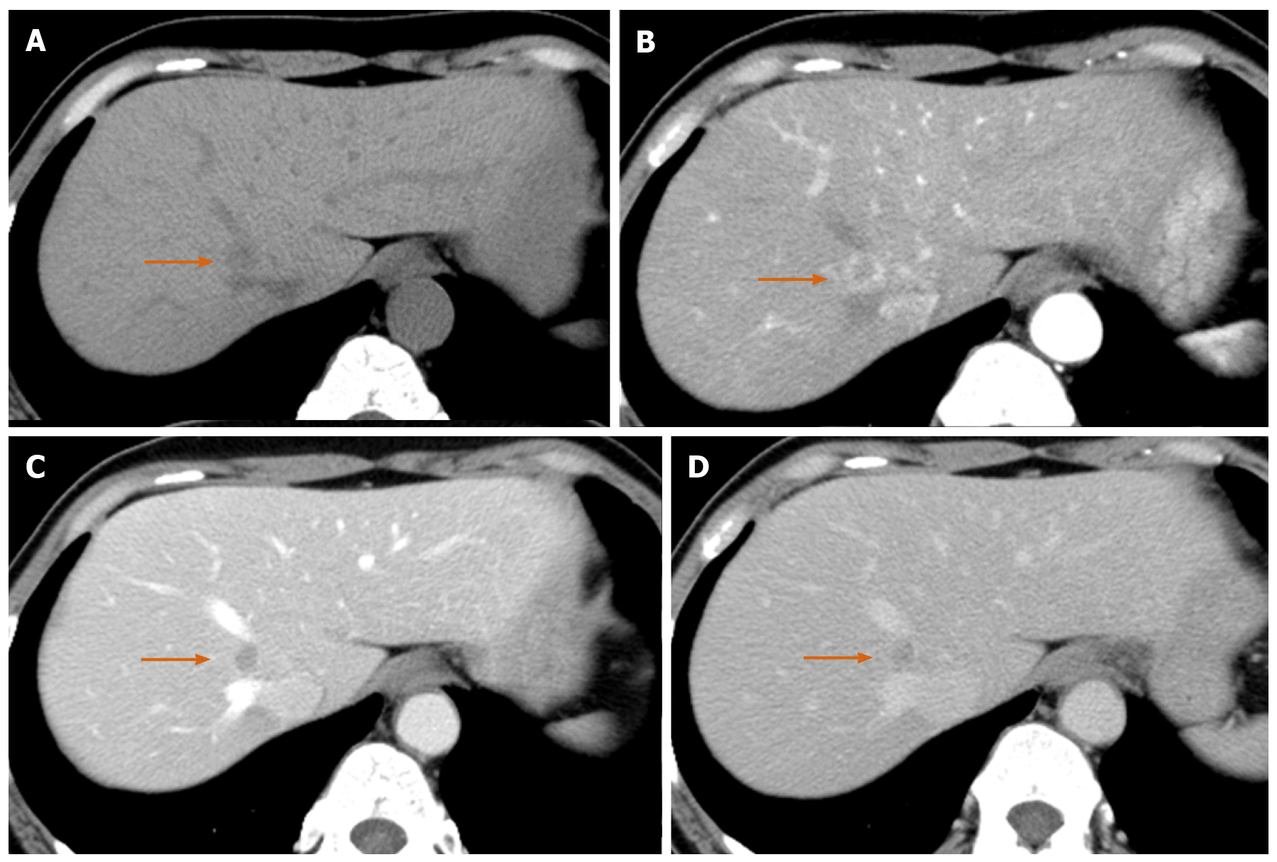
Figure 1 Computed tomography scan of a 54-year-old woman with reactive lymphoid hyperplasia (arrow).
A: Precontrast computed tomography showed a low density nodule in liver segment 1; B: The lesion showed perinodular enhancement in the arterial phase; C: The lesion showed washout in the portal phase; D: Although the lesion remained in low density in the equilibrium phase, the slight enhancement was observed in the lesion compared with the portal phase.
- Citation: Tanaka T, Saito K, Yunaiyama D, Matsubayashi J, Nagakawa Y, Tanigawa M, Nagao T. Diffusion-weighted imaging might be useful for reactive lymphoid hyperplasia diagnosis of the liver: A case report. World J Clin Cases 2020; 8(21): 5313-5319
- URL: https://www.wjgnet.com/2307-8960/full/v8/i21/5313.htm
- DOI: https://dx.doi.org/10.12998/wjcc.v8.i21.5313


