Copyright
©The Author(s) 2020.
World J Clin Cases. Nov 6, 2020; 8(21): 5304-5312
Published online Nov 6, 2020. doi: 10.12998/wjcc.v8.i21.5304
Published online Nov 6, 2020. doi: 10.12998/wjcc.v8.i21.5304
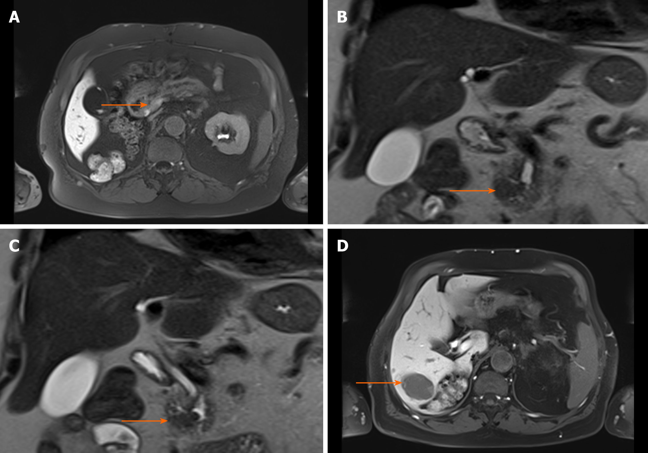
Figure 1 T2-weighted magnetic resonance images showing acinar cell carcinoma of the pancreatic head with synchronous liver metastases before surgery.
The patient’s presurgical bloodwork showed lipase of 5580 U/L, amylase of 172 U/L, C-reactive protein of 3.3 mg/dL, and leukocytes of 5 G/L. A: Primary tumor localized in the processus uncinatus; B and C: T2-weighted HASTE cor showing prepapillary tumor (arrow) infiltrating the common hepatic duct, which is extended; D: Hepatic metastasis in liver segment VI (arrow).
- Citation: Miksch RC, Schiergens TS, Weniger M, Ilmer M, Kazmierczak PM, Guba MO, Angele MK, Werner J, D'Haese JG. Pancreatic panniculitis and elevated serum lipase in metastasized acinar cell carcinoma of the pancreas: A case report and review of literature. World J Clin Cases 2020; 8(21): 5304-5312
- URL: https://www.wjgnet.com/2307-8960/full/v8/i21/5304.htm
- DOI: https://dx.doi.org/10.12998/wjcc.v8.i21.5304


