Copyright
©The Author(s) 2020.
World J Clin Cases. Nov 6, 2020; 8(21): 5296-5303
Published online Nov 6, 2020. doi: 10.12998/wjcc.v8.i21.5296
Published online Nov 6, 2020. doi: 10.12998/wjcc.v8.i21.5296
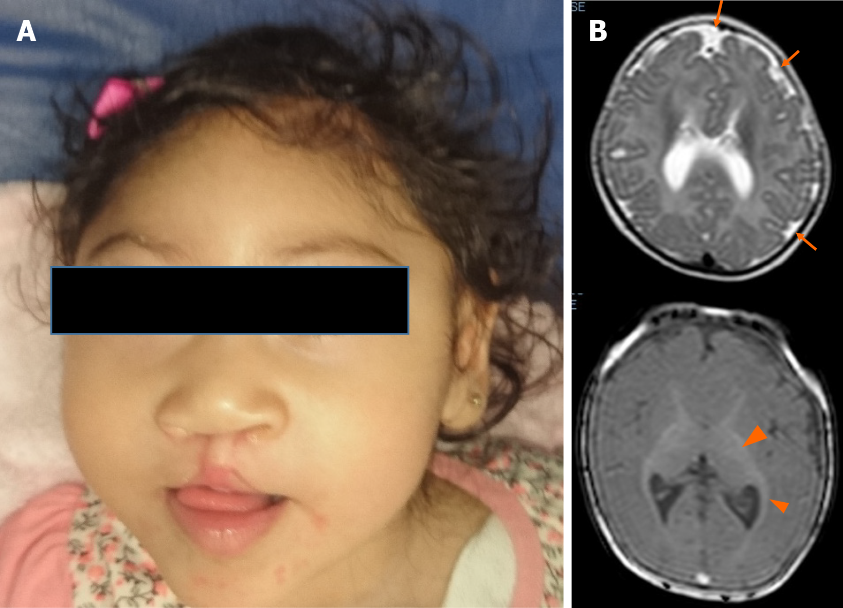
Figure 1 Patient at 1-year-old.
A: Microcephaly, upward-slanting palpebral fissures, depressed nasal bridge, bulbous nose, bilateral cleft lip, and palate are showed; B: The brain magnetic resonance scan (at five months) shows cortical atrophy, simplified gyral cortical patterns (orange arrow) and band heterotopia (arrow ahead).
- Citation: Toral-Lopez J, González Huerta LM, Messina-Baas O, Cuevas-Covarrubias SA. Submicroscopic 11p13 deletion including the elongator acetyltransferase complex subunit 4 gene in a girl with language failure, intellectual disability and congenital malformations: A case report . World J Clin Cases 2020; 8(21): 5296-5303
- URL: https://www.wjgnet.com/2307-8960/full/v8/i21/5296.htm
- DOI: https://dx.doi.org/10.12998/wjcc.v8.i21.5296


