Copyright
©The Author(s) 2020.
World J Clin Cases. Nov 6, 2020; 8(21): 5203-5212
Published online Nov 6, 2020. doi: 10.12998/wjcc.v8.i21.5203
Published online Nov 6, 2020. doi: 10.12998/wjcc.v8.i21.5203
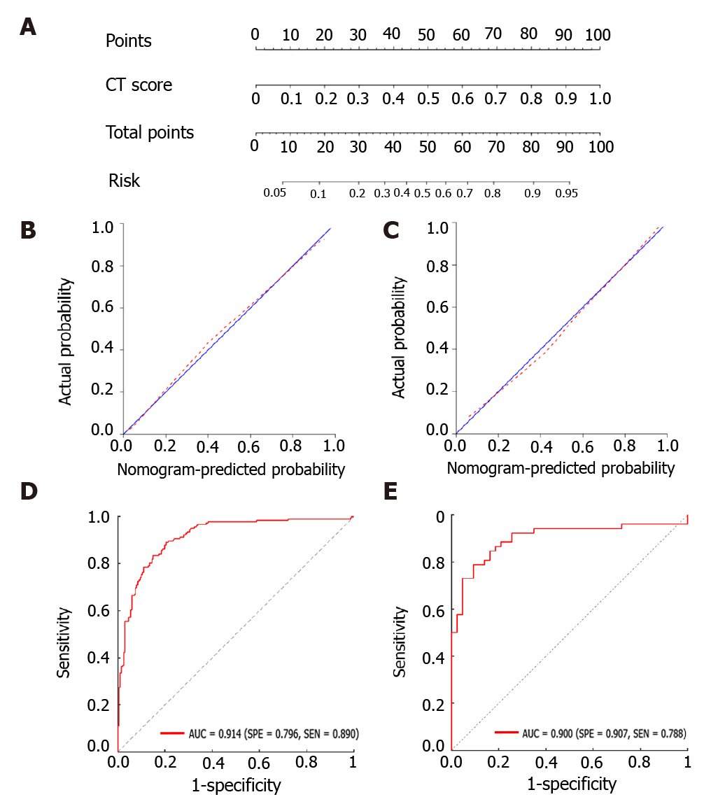
Figure 3 The radiomics nomogram for the differentiation of tuberculosis and lung cancer.
A: The construction of the nomogram model; B, C: The calibration curves of the nomogram model in the training group (B) and validation group (C), respectively; D, E: The receiver operating characteristic curves of the nomogram model in the training group (D) and validation group (E), respectively. CT: Computed tomography.
- Citation: Cui EN, Yu T, Shang SJ, Wang XY, Jin YL, Dong Y, Zhao H, Luo YH, Jiang XR. Radiomics model for distinguishing tuberculosis and lung cancer on computed tomography scans. World J Clin Cases 2020; 8(21): 5203-5212
- URL: https://www.wjgnet.com/2307-8960/full/v8/i21/5203.htm
- DOI: https://dx.doi.org/10.12998/wjcc.v8.i21.5203


