Copyright
©The Author(s) 2020.
World J Clin Cases. Nov 6, 2020; 8(21): 5159-5171
Published online Nov 6, 2020. doi: 10.12998/wjcc.v8.i21.5159
Published online Nov 6, 2020. doi: 10.12998/wjcc.v8.i21.5159
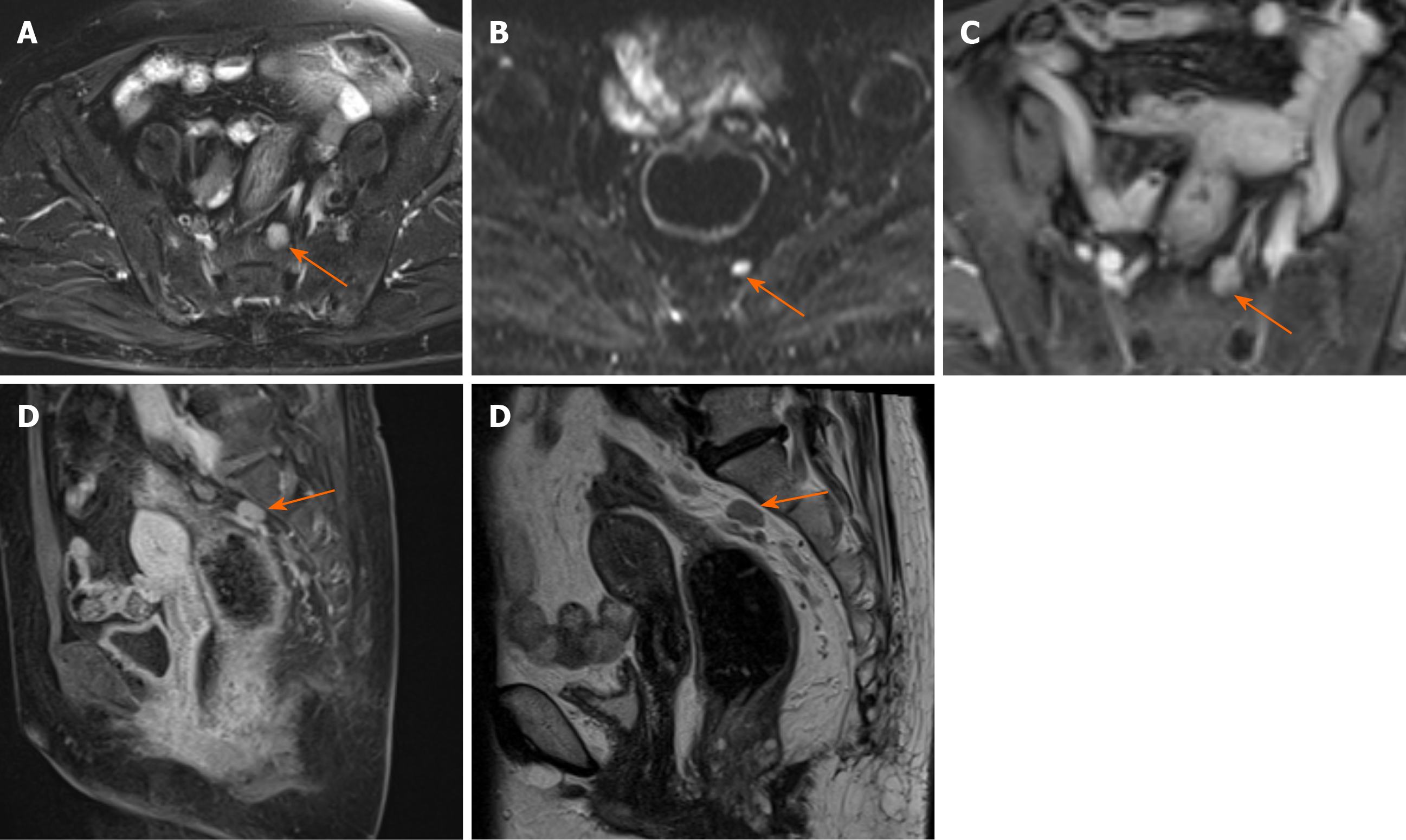
Figure 6 Magnetic resonance images of a 64-year-old woman with mucinous adenocarcinoma show perirectal lymph node with irregular morphology measuring > 5 mm in the short axis.
A: Fat-suppressed T2-weighted magnetic resonance imaging; B: Hyperintense on diffusion-weighted imaging; C and D: Heterogeneous enhancement on axial and sagittal dynamic contrast enhanced magnetic resonance imaging; E: Sagittal T2-weighted magnetic resonance imaging.
- Citation: Zhu X, Zhu TS, Ye DD, Liu SW. Magnetic resonance imaging findings of carcinoma arising from anal fistula: A retrospective study in a single institution. World J Clin Cases 2020; 8(21): 5159-5171
- URL: https://www.wjgnet.com/2307-8960/full/v8/i21/5159.htm
- DOI: https://dx.doi.org/10.12998/wjcc.v8.i21.5159


