Copyright
©The Author(s) 2020.
World J Clin Cases. Nov 6, 2020; 8(21): 5159-5171
Published online Nov 6, 2020. doi: 10.12998/wjcc.v8.i21.5159
Published online Nov 6, 2020. doi: 10.12998/wjcc.v8.i21.5159
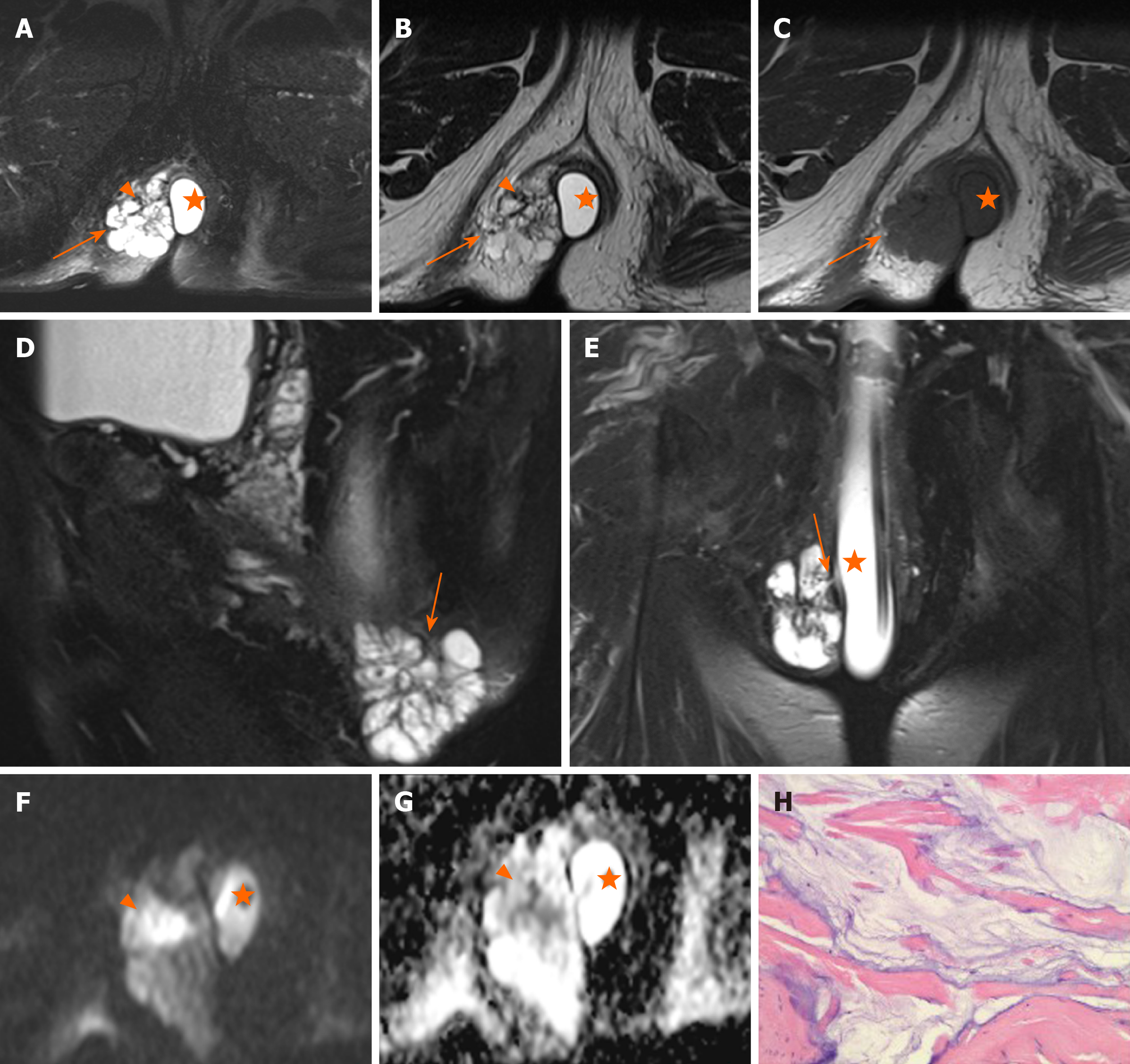
Figure 3 Magnetic resonance images of a 66-year-old man with mucinous adenocarcinoma.
A: Axial fat-suppressed T2-weighted magnetic resonance imaging (FS-T2WI); B: Axial T2WI; C: Axial T1-weighted magnetic resonance imaging; D: Sagittal FS-T2WI. A-D images show the tumor to the right. This lesion was multiloculated, cauliflower-like, and markedly hyperintense with focal slightly hypointense signal (orange triangle) on FS-T2WI, with a thin capsule (orange arrows) and focally unclear boundary; E: Coronal FS-T2WI shows a fistula between the mass and the anus; F: Axial diffusion-weighted image shows a slightly hyperintense with focal hyperintense lesion (orange triangle); G: Axial ADC map shows hyperintense with focal hypointense lesion (orange triangle). Artificial water sac can be seen in the rectum (orange star); H: hematoxylin and eosin staining (original magnification, ×200).
- Citation: Zhu X, Zhu TS, Ye DD, Liu SW. Magnetic resonance imaging findings of carcinoma arising from anal fistula: A retrospective study in a single institution. World J Clin Cases 2020; 8(21): 5159-5171
- URL: https://www.wjgnet.com/2307-8960/full/v8/i21/5159.htm
- DOI: https://dx.doi.org/10.12998/wjcc.v8.i21.5159


