Copyright
©The Author(s) 2020.
World J Clin Cases. Nov 6, 2020; 8(21): 5159-5171
Published online Nov 6, 2020. doi: 10.12998/wjcc.v8.i21.5159
Published online Nov 6, 2020. doi: 10.12998/wjcc.v8.i21.5159
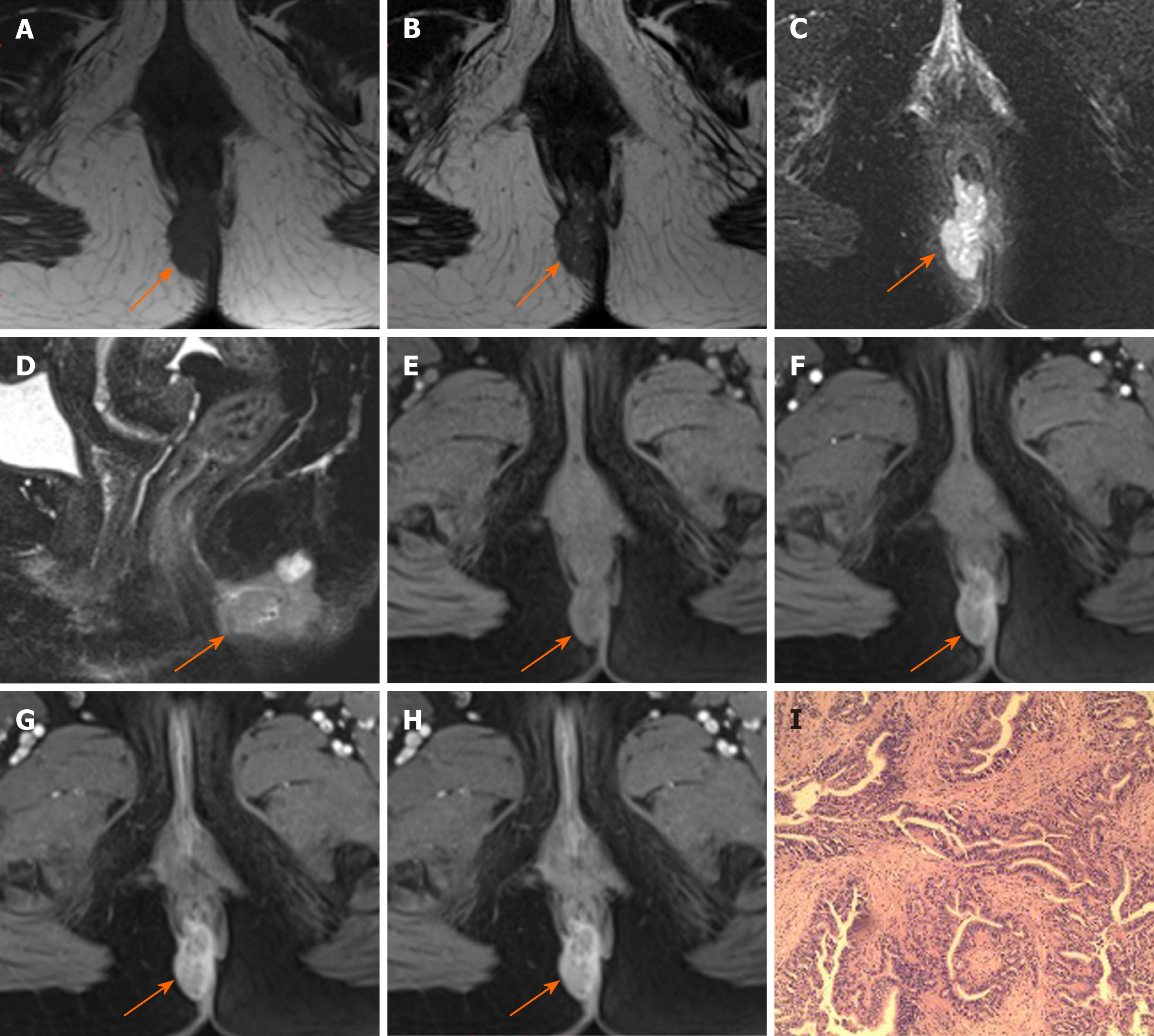
Figure 2 Magnetic resonance images of a 52-year-old woman with adenocarcinoma.
A: Axial T1-weighted magnetic resonance imaging; B: Axial T2WI; C: Axial fat-suppressed T2-weighted magnetic resonance imaging (FS-T2WI); D: Sagittal FS-T2WI images show the tumor to the right (orange arrows). The tumor is oval and hyperintense on FS-T2WI without capsule. E-H: Axial dynamic contrast-enhanced images show persistent heterogeneous enhancement (orange arrows); I: Hematoxylin and eosin stained image (original magnification, × 200).
- Citation: Zhu X, Zhu TS, Ye DD, Liu SW. Magnetic resonance imaging findings of carcinoma arising from anal fistula: A retrospective study in a single institution. World J Clin Cases 2020; 8(21): 5159-5171
- URL: https://www.wjgnet.com/2307-8960/full/v8/i21/5159.htm
- DOI: https://dx.doi.org/10.12998/wjcc.v8.i21.5159


