Copyright
©The Author(s) 2020.
World J Clin Cases. Oct 26, 2020; 8(20): 5042-5048
Published online Oct 26, 2020. doi: 10.12998/wjcc.v8.i20.5042
Published online Oct 26, 2020. doi: 10.12998/wjcc.v8.i20.5042
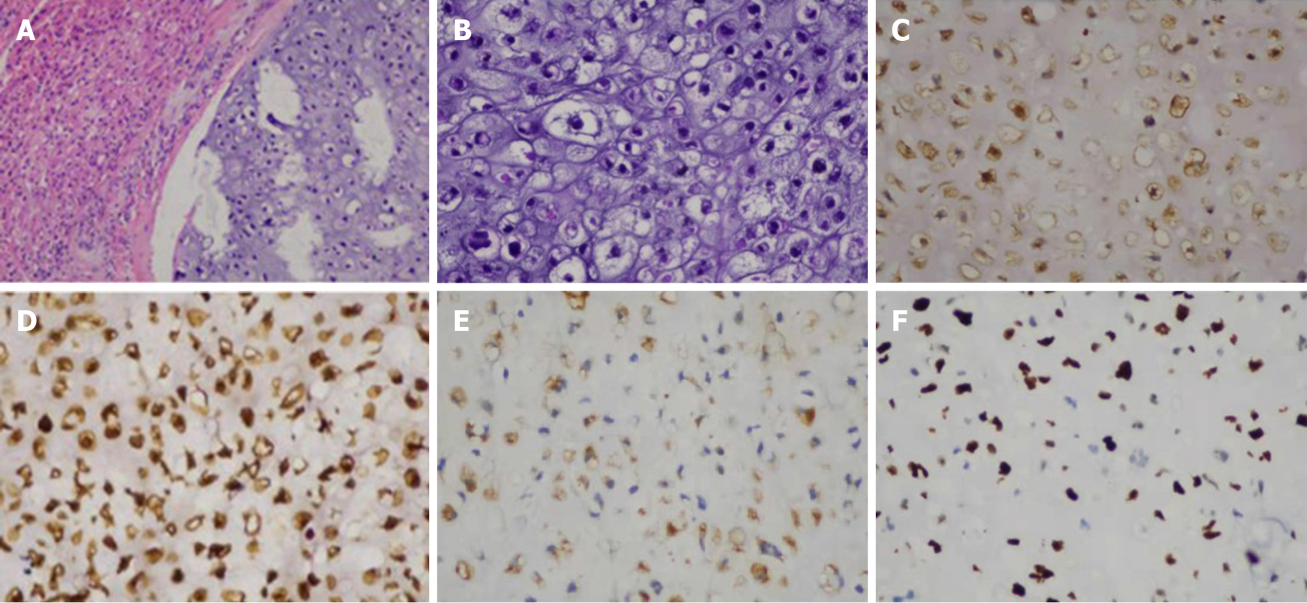
Figure 3 Histopathological evaluation.
A: Hematoxylin and eosin staining showed that tumor cells were unevenly distributed with cartilage pits, and the tumor was infiltrating and growing into surrounding liver tissue (magnifications 100 ×); B: Hematoxylin and eosin staining showed tumor cells with abnormal nuclei that were mostly mononuclear though occasionally binuclear (magnifications 200 ×); C: Tumor CK protein positive; D: Tumor vimentin protein positive; E: Tumor S-100 protein positive; F: Tumor Ki-67 immunostaining with 60% Ki-67 positive cells. (C-F immunohistochemistry, magnification 200 ×).
- Citation: Liu ZY, Jin XM, Yan GH, Jin GY. Primary chondrosarcoma of the liver: A case report. World J Clin Cases 2020; 8(20): 5042-5048
- URL: https://www.wjgnet.com/2307-8960/full/v8/i20/5042.htm
- DOI: https://dx.doi.org/10.12998/wjcc.v8.i20.5042


