Copyright
©The Author(s) 2020.
World J Clin Cases. Oct 26, 2020; 8(20): 5042-5048
Published online Oct 26, 2020. doi: 10.12998/wjcc.v8.i20.5042
Published online Oct 26, 2020. doi: 10.12998/wjcc.v8.i20.5042
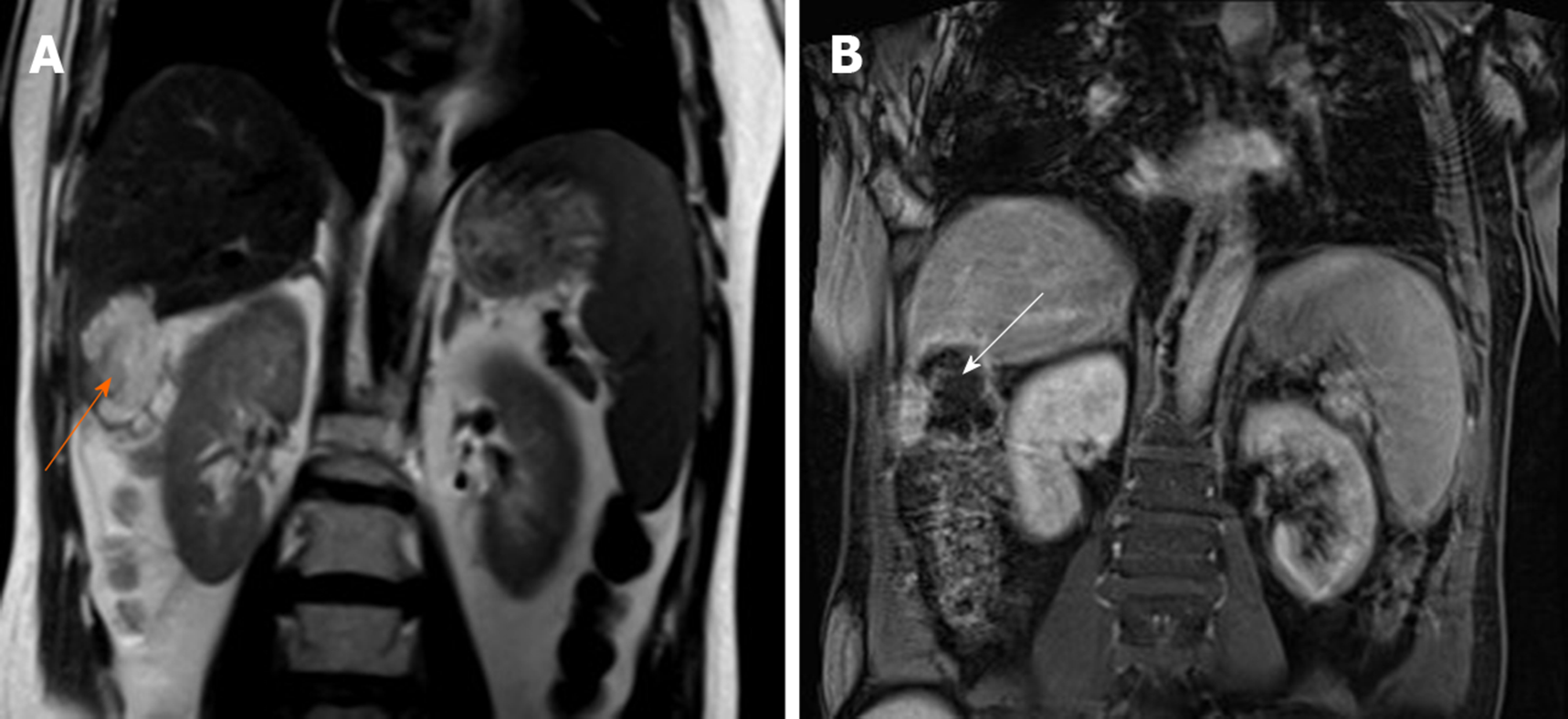
Figure 2 Magnetic resonance imaging after 3 mo.
A: A magnetic resonance T2 weighted image coronal image showed that the lesion was larger than before, and the border between the lesion and colonic liver curvature was unclear (orange arrow); B: A magnetic resonance T1 weight image coronal image (white arrow).
- Citation: Liu ZY, Jin XM, Yan GH, Jin GY. Primary chondrosarcoma of the liver: A case report. World J Clin Cases 2020; 8(20): 5042-5048
- URL: https://www.wjgnet.com/2307-8960/full/v8/i20/5042.htm
- DOI: https://dx.doi.org/10.12998/wjcc.v8.i20.5042


