Copyright
©The Author(s) 2020.
World J Clin Cases. Oct 26, 2020; 8(20): 5019-5024
Published online Oct 26, 2020. doi: 10.12998/wjcc.v8.i20.5019
Published online Oct 26, 2020. doi: 10.12998/wjcc.v8.i20.5019
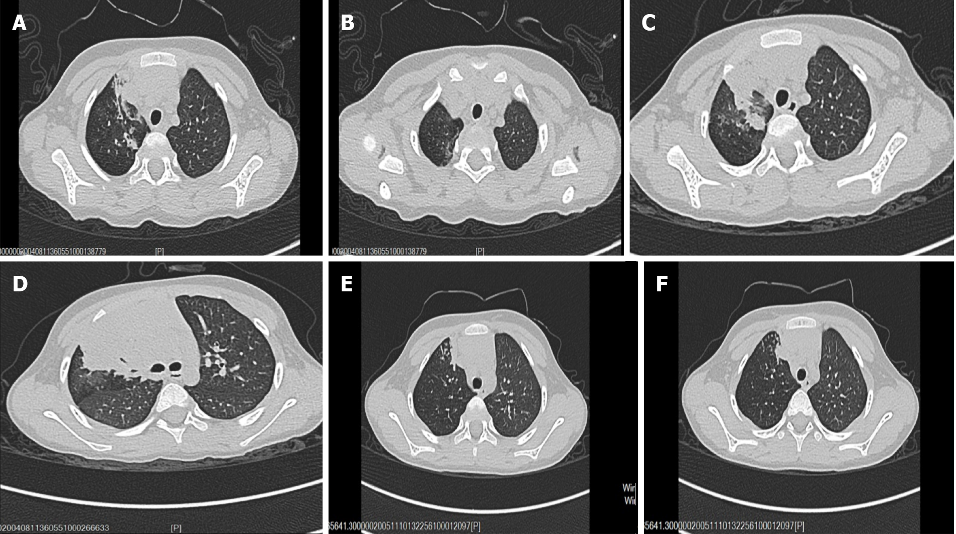
Figure 1 Radiological findings.
A and B: Chest computed tomography (CT) images showing right upper lobe pneumonia with segmental atelectasis; C and D: Chest CT images showing that the inflammatory lesions of the right upper lung were expanded and new lesions appeared in the right lower lung; E and F: Chest CT images showing that the inflammatory lesions were diminished and the atelectasis was partially restored.
- Citation: Liu YR, Ai T. Plastic bronchitis associated with Botrytis cinerea infection in a child: A case report. World J Clin Cases 2020; 8(20): 5019-5024
- URL: https://www.wjgnet.com/2307-8960/full/v8/i20/5019.htm
- DOI: https://dx.doi.org/10.12998/wjcc.v8.i20.5019


