Copyright
©The Author(s) 2020.
World J Clin Cases. Oct 26, 2020; 8(20): 4993-4998
Published online Oct 26, 2020. doi: 10.12998/wjcc.v8.i20.4993
Published online Oct 26, 2020. doi: 10.12998/wjcc.v8.i20.4993
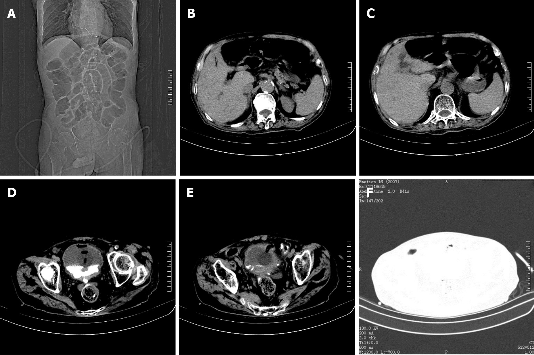
Figure 1 Computed tomography image of the patient’s abdomen before surgery.
A: The catheter was indwelling, and the intestinal cavity was dilated; B: A small amount of free gas in the abdominal cavity; C: A small amount of effusion around the liver and spleen; D and F: Air collected in the bladder; E: The bladder wall was thickened, and the end of catheter was sighted in the bladder wall.
- Citation: Wu B, Wang J, Chen XJ, Zhou ZC, Zhu MY, Shen YY, Zhong ZX. Bladder perforation caused by long-term catheterization misdiagnosed as digestive tract perforation: A case report. World J Clin Cases 2020; 8(20): 4993-4998
- URL: https://www.wjgnet.com/2307-8960/full/v8/i20/4993.htm
- DOI: https://dx.doi.org/10.12998/wjcc.v8.i20.4993


