Copyright
©The Author(s) 2020.
World J Clin Cases. Oct 26, 2020; 8(20): 4975-4980
Published online Oct 26, 2020. doi: 10.12998/wjcc.v8.i20.4975
Published online Oct 26, 2020. doi: 10.12998/wjcc.v8.i20.4975
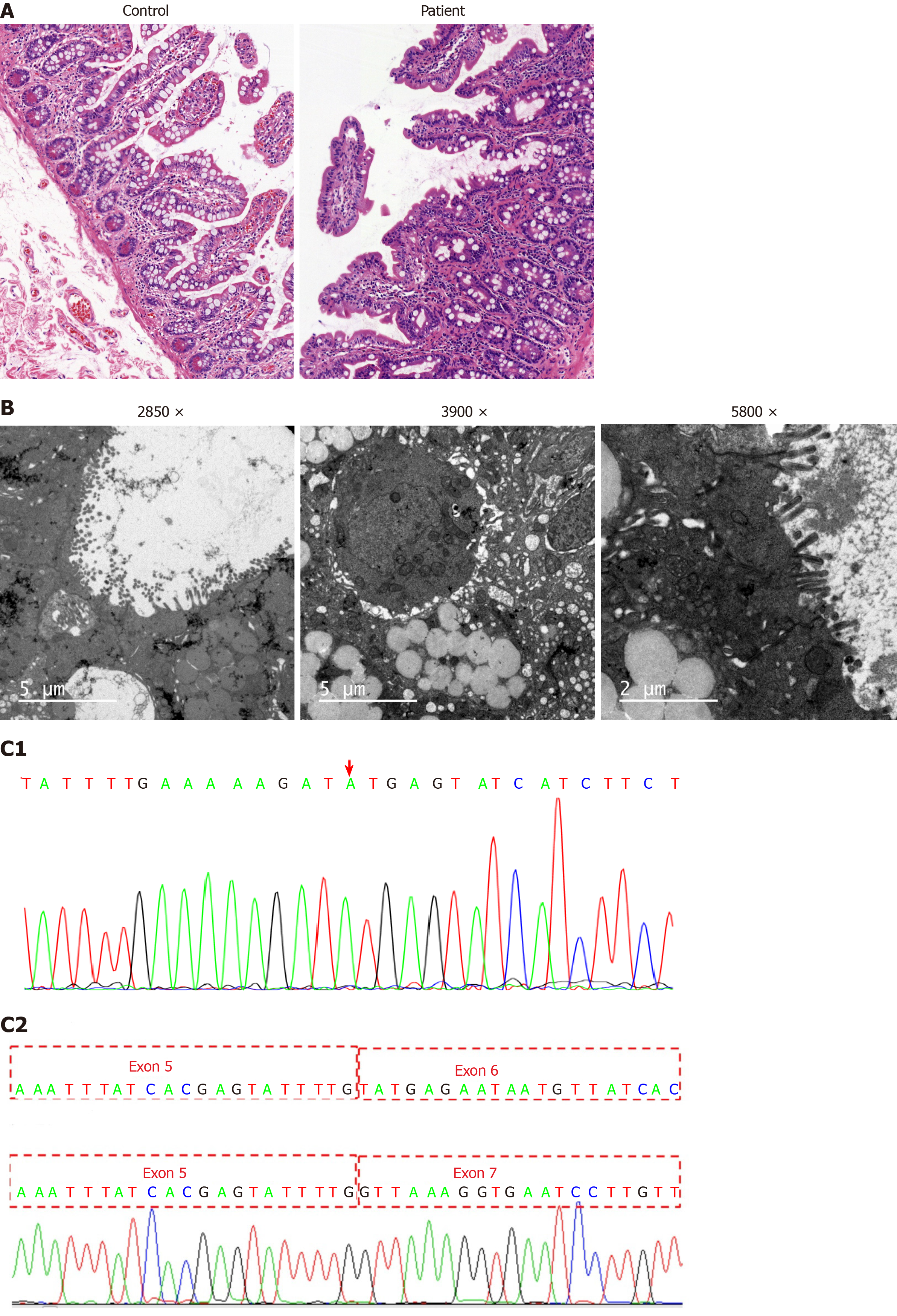
Figure 1 Histology and whole-exome sequencing and analysis.
A: Histopathological findings by light microscopy: Hematoxylin and eosin staining revealed enterocytes with focal resembling tufts in the patient’s jejunal biopsies (healthy mother was used as a control); B: Histopathological findings by electron microscope: Electric mirror reports microvilli sparse and vacuolar degeneration of epithelial cells; C1: Whole-exome sequencing showing a novel homozygous splice mutation (c.657+1[IVS6]G>A) in the epithelial cell adhesion molecule gene in the patient; C2: Sequence analysis results showing the loss of exon 6 through mutation.
- Citation: Zhou YQ, Wu GS, Kong YM, Zhang XY, Wang CL. New mutation in EPCAM for congenital tufting enteropathy: A case report. World J Clin Cases 2020; 8(20): 4975-4980
- URL: https://www.wjgnet.com/2307-8960/full/v8/i20/4975.htm
- DOI: https://dx.doi.org/10.12998/wjcc.v8.i20.4975


