Copyright
©The Author(s) 2020.
World J Clin Cases. Oct 26, 2020; 8(20): 4966-4974
Published online Oct 26, 2020. doi: 10.12998/wjcc.v8.i20.4966
Published online Oct 26, 2020. doi: 10.12998/wjcc.v8.i20.4966
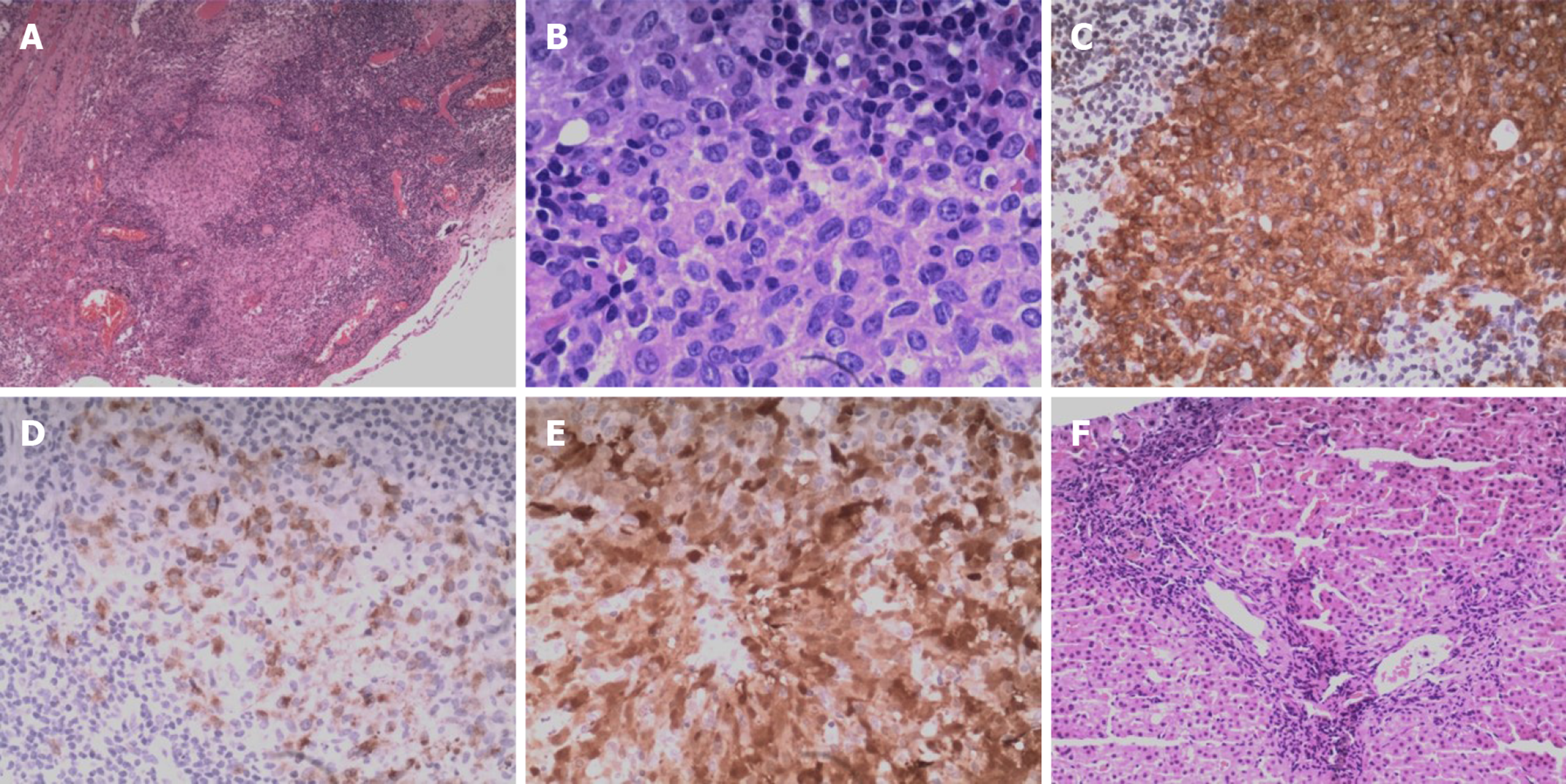
Figure 4 Histologic examination of the right pulmonary bullae sections and liver biopsy samples.
A: Nodular clusters of histiocytes were detectable upon histologic assessment of the bullae samples, as was evidence of fibrosis and inflammation (hematoxylin & eosin, × 4). B: The cells in bullae samples exhibited a pronounced cytoplasm as well as mild cytologic atypia, slight nuclear enlargement, and the presence of nuclear grooves (hematoxylin & eosin, × 40). C-E: Langerhans cells show immunopositivity for CD1a (C), CD207 (D), and S-100 (E) (× 20). F: Liver biopsy demonstrated chronic portal fibrosis with ductular damage and proliferation (hematoxylin & eosin, × 10).
- Citation: Wang BB, Ye JR, Li YL, Jin Y, Chen ZW, Li JM, Li YP. Multisystem involvement Langerhans cell histiocytosis in an adult: A case report. World J Clin Cases 2020; 8(20): 4966-4974
- URL: https://www.wjgnet.com/2307-8960/full/v8/i20/4966.htm
- DOI: https://dx.doi.org/10.12998/wjcc.v8.i20.4966


