Copyright
©The Author(s) 2020.
World J Clin Cases. Oct 26, 2020; 8(20): 4966-4974
Published online Oct 26, 2020. doi: 10.12998/wjcc.v8.i20.4966
Published online Oct 26, 2020. doi: 10.12998/wjcc.v8.i20.4966
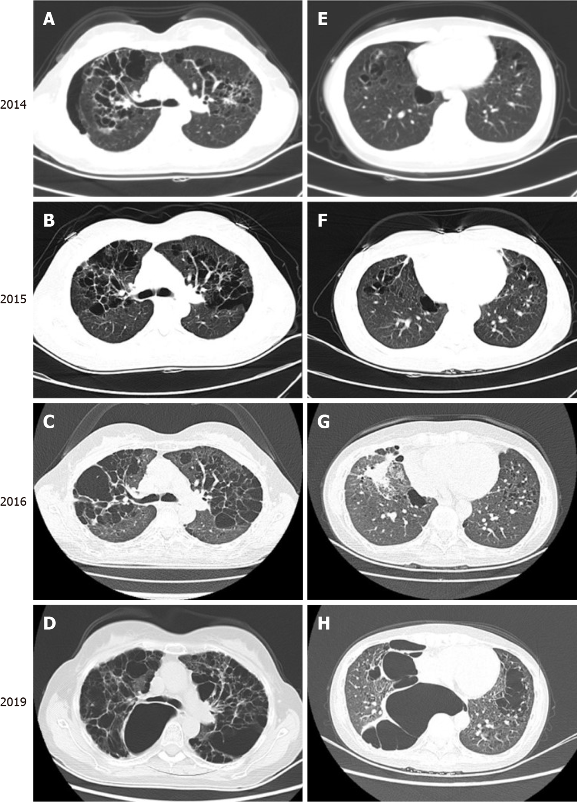
Figure 3 Chest computed tomography findings over a 5-year period.
Computed tomography results from January 2014 (A and E), January 2015 (B and F), September 2016 (C and G), and July 2019 (D and H) record the growth and progression of multiple thinly-walled cystic lesions over 5 years, predominantly in the upper and middle lungs, with some lower lung involvement. These cysts were of variable size, and some were confluent with one another.
- Citation: Wang BB, Ye JR, Li YL, Jin Y, Chen ZW, Li JM, Li YP. Multisystem involvement Langerhans cell histiocytosis in an adult: A case report. World J Clin Cases 2020; 8(20): 4966-4974
- URL: https://www.wjgnet.com/2307-8960/full/v8/i20/4966.htm
- DOI: https://dx.doi.org/10.12998/wjcc.v8.i20.4966


