Copyright
©The Author(s) 2020.
World J Clin Cases. Oct 26, 2020; 8(20): 4966-4974
Published online Oct 26, 2020. doi: 10.12998/wjcc.v8.i20.4966
Published online Oct 26, 2020. doi: 10.12998/wjcc.v8.i20.4966
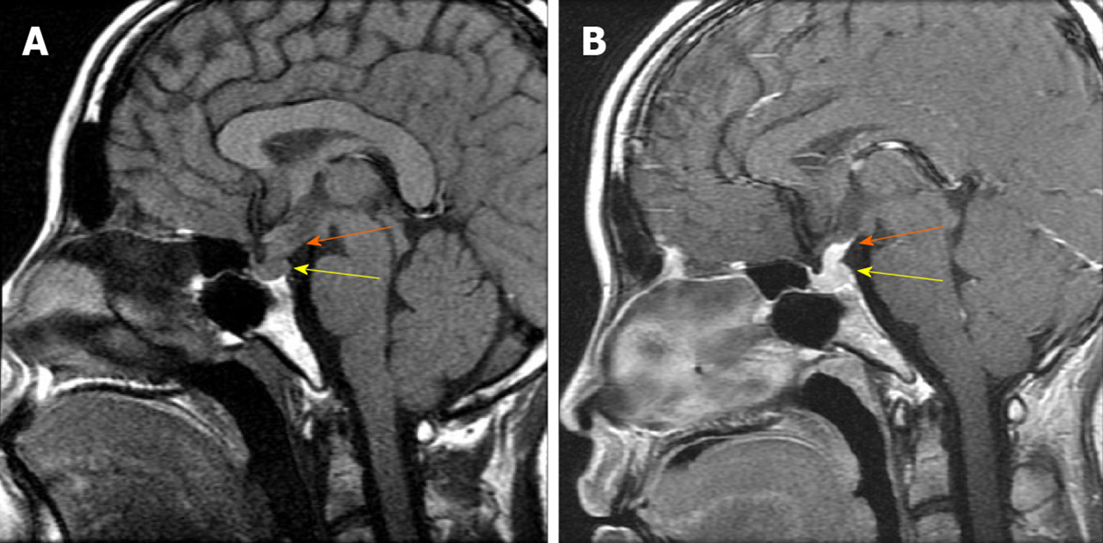
Figure 1 Pituitary magnetic resonance imaging findings.
A: T1-weighted magnetic resonance imaging (MRI) images from May 2007 reveal posterior pituitary lesions (yellow arrow) with the absence of the posterior ‘bright spot’ and an enlarged pituitary stalk (orange arrow), whereas the anterior pituitary, supra sellar cistern, and adjacent skull were normal; B: Enhanced T1-weighted images (WIs) MRI images from May 2007 show uniform intensification of the posterior pituitary (yellow arrow) and the enlarged pituitary stalk (orange arrow).
- Citation: Wang BB, Ye JR, Li YL, Jin Y, Chen ZW, Li JM, Li YP. Multisystem involvement Langerhans cell histiocytosis in an adult: A case report. World J Clin Cases 2020; 8(20): 4966-4974
- URL: https://www.wjgnet.com/2307-8960/full/v8/i20/4966.htm
- DOI: https://dx.doi.org/10.12998/wjcc.v8.i20.4966


