Copyright
©The Author(s) 2020.
World J Clin Cases. Oct 26, 2020; 8(20): 4883-4894
Published online Oct 26, 2020. doi: 10.12998/wjcc.v8.i20.4883
Published online Oct 26, 2020. doi: 10.12998/wjcc.v8.i20.4883
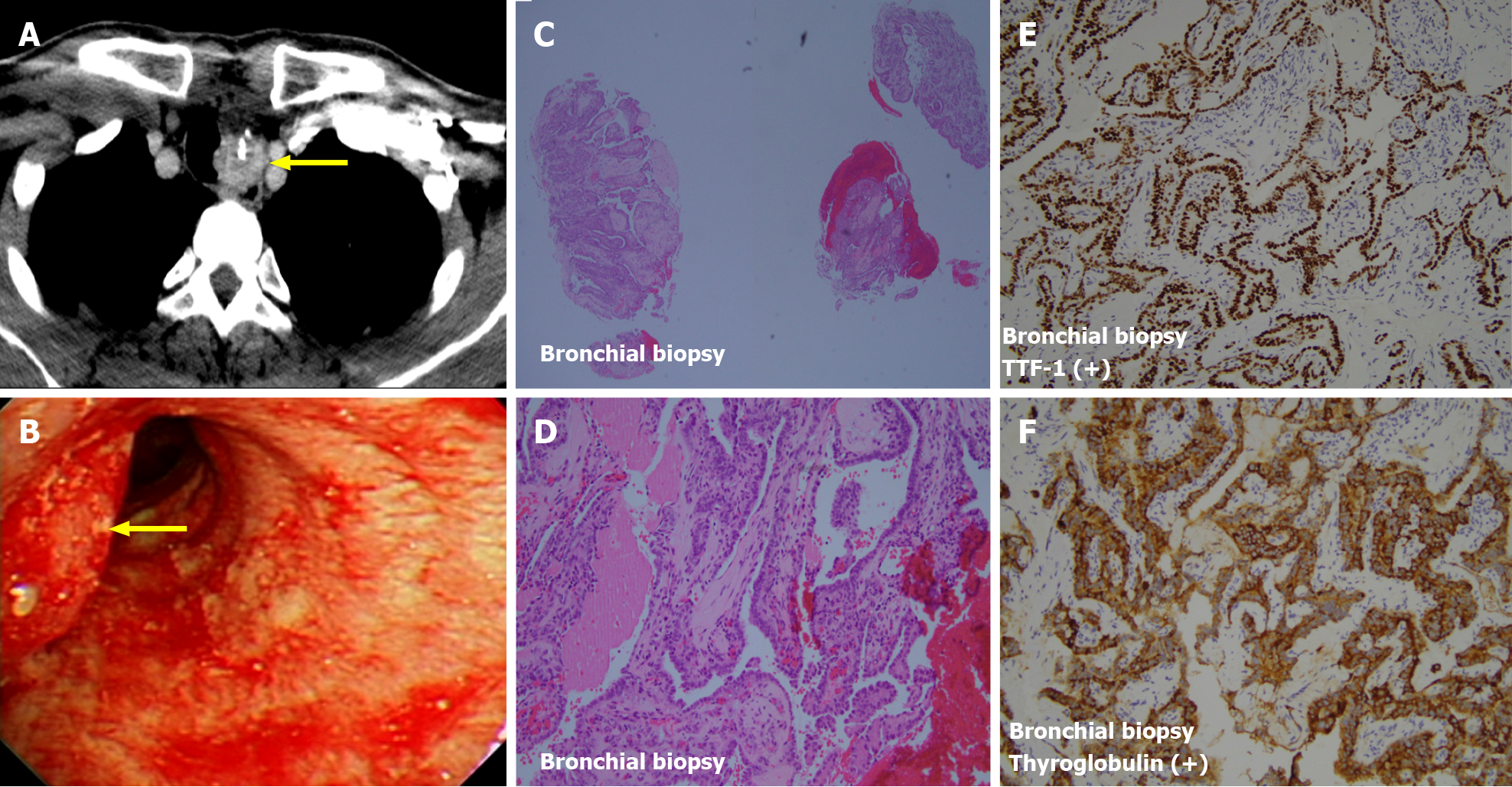
Figure 2 Computed tomography images and staining results.
A and B: Left para-tracheal lesion was first observed on computed tomography (A) and directly visualized under bronchoscopy (B); C and D: Biopsy was conducted, and it showed pictures of neoplastic cells growth in papillary pattern with atypical large vesicular and pale nuclei with obvious nuclear groove; E and F: It was stained with TTF-1 100% (E) and thyroglobulin 100% (F).
- Citation: Yang CH, Chen KT, Lin YS, Hsu CY, Ou YC, Tung MC. Improvement of lenvatinib-induced nephrotic syndrome after adaptation to sorafenib in thyroid cancer: A case report. World J Clin Cases 2020; 8(20): 4883-4894
- URL: https://www.wjgnet.com/2307-8960/full/v8/i20/4883.htm
- DOI: https://dx.doi.org/10.12998/wjcc.v8.i20.4883


